Lecture
The most natural approach to the development of a model of the human visual system would apparently be a physiological analysis of the eye, the nerve pathways from the eye to the brain, and those parts of the brain that are associated with visual perception. Unfortunately, we are not able to solve this problem due to the fact that the visual system consists of a huge number of very small elements. However, as a result of physiological studies of the eye, many data have already been obtained that are useful for creating a model of the visual system [4-7].
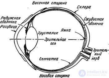
In fig. 2.2.1 shows a cross section of a human eyeball. The front transparent outer shell of the eye is called the cornea. The rest of the outer shell, the sclera, consists of dense fibers. The next layer is the choroid, which contains capillaries that supply blood to the eye. Inside the choroid is the retina with receptors of two types - rods and cones. The nerve fibers connected to the retina emerge from the eyeball in the form of a bundle - the optic nerve. The light that enters the eye through the cornea focuses on the surface of the retina with a lens, the shape of which changes under the influence of a special muscle to ensure good focusing of objects at different distances from the eye. The iris acts as a diaphragm, changing the amount of light that passes into the eye.

Fig. 2.2.1. Cross-section of the eye.
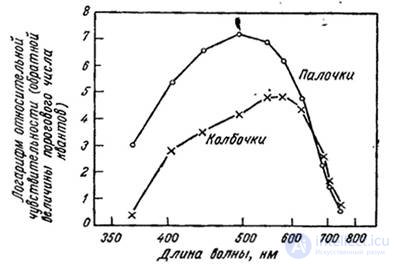
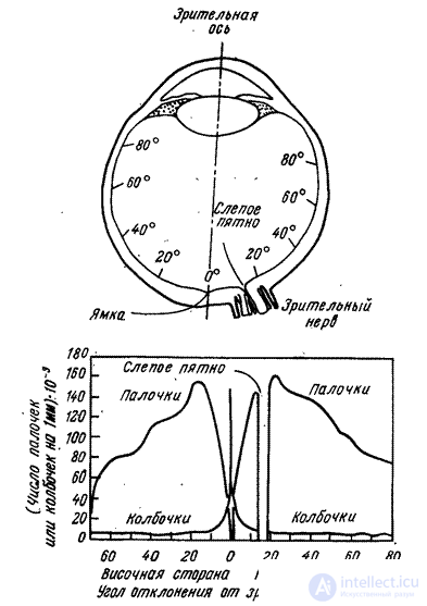
The rods are long, thin receptors, and cones are shorter and thicker. These receptors act in different ways. Rods are more sensitive to light than cones. In case of low illumination, the rods provide the reaction of the visual system (night vision). Cones function in high light conditions, their reaction represents day vision. In fig. 2.2.2 shows the relative sensitivity of rods and cones, depending on the wavelength of the perceived light [7, 8]. The eye contains about 6.5 million cones and 100 million rods. The distribution of rods and cones over the retina is shown in fig. 2.2.3 (4 1. The greatest density of cones falls on a small area of the retina called the central fossa which is near the exit from the eye of the optic nerve. This is the region of the sharpest day vision. In the vicinity of the optic nerve there are no rods or cones - this is a blind spot of the eye .
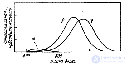
In recent years, it has been established that there are three main types of retinal cones [9, 10). These cones have different spectral characteristics of light absorption with a maximum in the red, green and blue regions of the optical spectrum. In fig. 2.2.4 shows the spectral absorption curves of retinal pigments [10]. Two features of these curves should be noted: first, the relatively low sensitivity of type a cones, which perceive mainly blue light, and, second, a significant overlap of the curves. The existence of three types of cones serves as the physiological basis for the three-color theory of color vision.

Fig. 2.2.2. Sensitivity of rods in cones according to Vald [7, 8].
When the light excites a stick or cone, a photochemical transition occurs, resulting in a nervous impulse. The mechanism of propagation of nerve impulses in the visual system is currently not fully understood. It is known that the optic nerve contains about 800,000 nerve fibers. The retina has over 100 million receptors. Since photochemical processes in the retina and the mechanism of propagation of nerve impulses in the eye are not well understood, it is impossible to give a complete description of the visual processes. We have to be satisfied with the development of models that describe and, hopefully, predict the reaction of the human visual system to certain images. The following section describes some of the phenomena that should be considered when modeling the visual system.

Fig. 2.2.3. Distribution of rods and cones over the retina [4].

Fig. 2.24. Spectral absorption curves of retinal receptor pigments [10]
Comments
To leave a comment
Digital image processing
Terms: Digital image processing