
Lecture
Это продолжение увлекательной статьи про депрессия.
...
depression is no higher than that resulting from antidepressant use, and the risk of suicide is not increased .
Since the late 1990s and early 2000s, new approaches to the treatment of depression have been studied, such as repetitive transcranial magnetic stimulation, vagus nerve stimulation, magnetic seizure therapy, and deep brain stimulation. Their antidepressant effect has for the most part not yet been sufficiently proven, and yet these methods are already used in some countries: transcranial magnetic stimulation — in Canada and Israel, vagus nerve stimulation — in the United States as an adjunctive method in the treatment of depression .
Data on the effectiveness of transcranial magnetic stimulation (TMS) are limited and contradictory . There is evidence showing its effectiveness compared to ECT, as well as its effectiveness when combined with certain psychopharmacological agents . The side effects and long-term changes in brain functioning associated with repetitive TMS have been little studied , but it is known that TMS is characterized, in particular, by a risk of switching into hypomanic/manic states (especially in patients with bipolar affective disorder) and a risk of psychotic symptoms manifesting .
A more advanced version of transcranial magnetic stimulation is the FDA-approved method of Deep Transcranial Magnetic Stimulation (Deep TMS), approved for the treatment of severe forms of depression (along with other conditions).
Vagus nerve stimulation is approved by the FDA in the United States as an adjunctive treatment for the long-term therapy of chronic or recurrent depression in patients who have not adequately responded to 4 or more properly selected antidepressants. Data on the antidepressant activity of this method are limited. Because of the invasive nature of this method, its use must be limited to exceptional clinical cases where there is a high need for such treatment, other interventions have failed, and a clinical effect is likely .
Narcotherapy (medical sleep therapy) has low evidentiary support in the treatment of depression; efferent methods (plasmapheresis, extracorporeal pharmacotherapy, ultraviolet therapy, laser therapy), hyperbaric oxygenation, and craniocerebral hypothermia have an even lower degree of evidentiary support . Hemodialysis, franklinization (right-sided and left-sided), and low-frequency alternating magnetic field therapy may also be used , as well as microwave resonance therapy and lateral therapy[55]. An extremely low level of evidence characterizes clinical studies of such treatments for depression as manual acupuncture, aromatherapy, biofeedback, herbal medicine, homeopathy, hypnosis, massage, dance therapy, qigong, yoga, comprehensive diets, fasting, and the use of inositol, magnesium, omega-3 fatty acids, probiotics, tryptophan, vitamins B and D, and zinc . These are for the most part auxiliary (complementary) methods that can largely be used alongside rational pharmacotherapy, without replacing it.
It has been shown that people suffering from depression often have lower omega-3 reserves in their bodies than those who are not depressed; and the scarcer this reserve, the more severe the symptoms. The amount of omega-3 in the daily diet affects the tendency to develop depression. Studies suggest that in some cases an antidepressant effect can be achieved by taking a certain daily amount of a mixture of two types of fish oil: eicosapentaenoic acid and docosahexaenoic acid, preferably combined with vitamins E and C and selenium . In 4 of 7 double-blind RCTs, depressive symptoms were shown to be significantly reduced with regular injections of eicosapentaenoic acid as an adjunct to antidepressant treatment, though other data cast doubt on whether this type of therapy has a genuine antidepressant effect on its own .
Depression can also be treated with music therapy, art therapy , occupational therapy, color therapy , animal-assisted therapy, balneotherapy[99], oil baths[111]:186, and magnetic field exposure to the patient's brain (magnetotherapy). These are likewise adjunctive methods that are not advisable to use as monotherapy.
Autogenic training may also be used — a method of relieving tension through one's own efforts, carried out individually or in a group. However, this type of treatment is not applicable in acute depression, since the patient is incapable of the concentration required for it and unable to relax. Autogenic training can be beneficial once the patient's condition has already improved; it can help relieve individual symptoms of depression, such as insomnia and headache caused by muscle spasms, and help ease the anxiety felt by the patient[111]:186—187.
There are suggestions of possible efficacy of dosed hypoxia, meditation, and neurosurgical methods[33].
In the 1950s—60s, studies of psychedelics in the treatment of depression were also conducted in the USA and the USSR[122].
Psychoses occur rarely during pregnancy, but at childbirth they are observed 10 times more often than at other periods of a woman's life. The fact that in most cases these are depressive psychoses finds its explanation in the clinical picture. In terms of symptomatology they partly correspond to melancholias (of course, reactive depressions also occur during the postpartum period), and are partly presented as atypical or schizoaffective syndromes (incidentally, schizophrenia also arises during the postpartum period). Postpartum depressions mostly begin in the first or second week after childbirth. Some of these women subsequently remain healthy, while about 1/3 fall ill with psychoses again.
No link has been established between the climacteric, or menopause, and melancholia. Melancholias also arise at this age, either as recurrent phases or as first manifestations, and biological and psychological processes during the climacteric may serve as a provoking factor. A statistical accumulation of melancholias during the climacteric period is not confirmed.
More often what is involved are non-psychotic psychovegetative syndromes with such well-known symptoms as hot flashes, bouts of sweating, tachycardia, dizziness, and, in part, depressive mood. This climacteric syndrome is multifactorial in origin. Alongside hormonal factors, one must take into account the situational problems of this period of bodily reorganization and premorbid factors. Therapeutically, hormones are indicated only in the most severe cases; psychopharmacological agents are always useful; psychotherapeutic assistance in working through the conflict is necessary.
The psychopathology arising in traumatic brain injuries confirms the role of the temporal, as well as the frontal, lobes of the brain in the development of depression.
Hypothymia in the acute period of concussion
Affective (subaffective) disorders are found in 68% of patients in the acute period after concussion and are characteristic of neurotic-level disorders. Hypothymia is the most fully represented condition in the psychopathology of the acute period of concussion and occurs in 50% of cases. Anxious subdepression (36% of patients in the acute period of concussion) and asthenic subdepression (11%) are most commonly observed.
How can a person help themselves? It is a mistaken belief among patients that carefully taking medications and attending psychotherapy sessions can quickly eliminate depression. Active participation of patients in the treatment process itself is extremely necessary.
Take care of yourself, be a little selfish. Give yourself a bit more time, and relax.
Always get enough sleep. Go to bed at the same time, and keep the room shielded from daylight, as this promotes the production of melatonin (the sleep hormone).
Lead an active lifestyle. Movement is the best antidepressant. Daily walks in the fresh air give the body energy and help maintain a good mood. Physical exercise or any physical activity helps lift your mood, and not simply because it distracts you from your problems — muscle tone also increases. Vigorous exercise causes the brain to release natural substances — endorphins. These substances are produced by the human body and are similar in their action to antidepressants and antipsychotics. In addition, exercise makes you treat yourself better, and when you treat yourself better, you gain new strength to continue the fight against depression.
Eat healthy food. Think about your diet, and make sure your food is low in fat, high in protein, and rich in vitamins and minerals. Some foods contain natural ingredients that help prevent mood decline and help restore well-being.
Communicate with friends and acquaintances. Depression isolates you, shutting you within walls of grief. If you feel that you are not communicative, or worry that your acquaintances or friends have distanced themselves from you and think badly of you, try talking to them about it. When we feel bad, our judgments about others change. Whenever possible, try to maintain your previous contacts with friends and colleagues.
Techniques that encourage creativity, self-disclosure, and understanding of underlying problems (music therapy, color therapy, art therapy, aromatherapy, dance therapy) are very helpful in preventing the illness, and they are also very effectively used in psychotherapy.
Do not use psychotropic and psychoactive substances without first consulting a doctor. Alcohol, drugs, and sedatives will bring only temporary relief and will cause dependence.
If you have problems you cannot cope with, seek the help of a psychotherapist. This will allow you to take timely measures.
Depression (from the Latin Deprimo — to press down) — is a mental illness characterized by the «depressive triad», which includes the following disturbances:
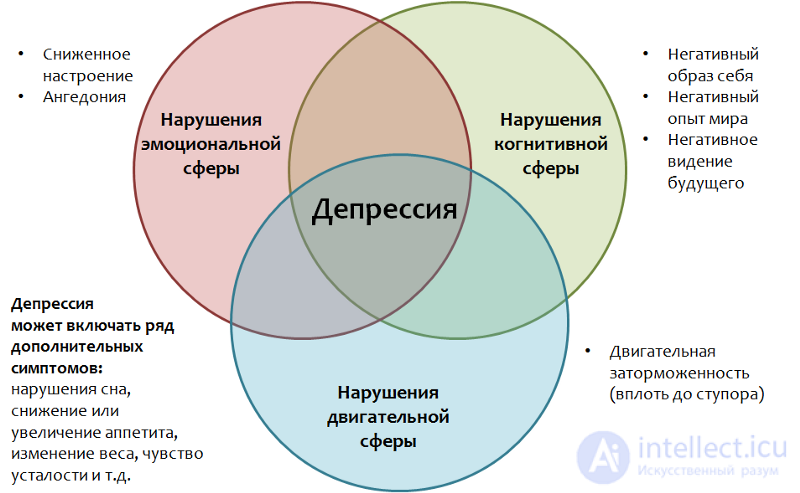 Psychiatry: a national guide. Moscow: GEOTAR-Media, 2009. 1000 p.
There are also additional symptoms: loss or increase of appetite (loss or gain of weight), sleep disturbances (drowsiness or insomnia), a feeling of fatigue, increased tiredness, etc.
According to WHO data (Fact sheet No. 369), more than 300 million people suffer from depression, and this figure tends to increase. Depression differs from ordinary mood changes and short-term emotional reactions to problems in everyday life. It can lead to serious health impairments, social maladjustment, and reduced capacity for work and learning. In the worst cases, it can lead to suicide.
Approximately 800,000 people die by suicide every year — the second leading cause of death among people aged 15–29.
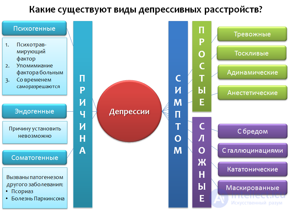 Fig. 1 Depression: The Treatment and Management of Depression in Adults (Updated Edition). Leicester (UK): British Psychological Society, 2010.
Let's look at a simplified classification of depressive disorders (Fig. 1), which is not exhaustive but includes the main examples. Depressions can be classified by their cause (etiology) and by the predominance of particular symptoms, as well as by various combinations of these.
In the first case, three categories can be distinguished:
By predominant symptom, depressions can be: anxious, melancholic, adynamic, anesthetic (experiencing an absence of emotions — «emotional anesthesia») — these are examples of «simple depressions».
«Complex depressions» combine symptoms of depression with other psychopathologies: depression with delusions, hallucinations, catatonic depression, masked depression — where symptoms are disguised as diseases of internal organs, or «somatized» — headaches, abdominal pain, chest pain, etc.
The difficulty in treating depressive disorders lies in the absence of complete recovery in severe or complicated forms of depressive disorders. However, self-resolution of the condition is also possible if the course is not severe and there are no complications in the form of symptoms of other psychopathologies, and so on.
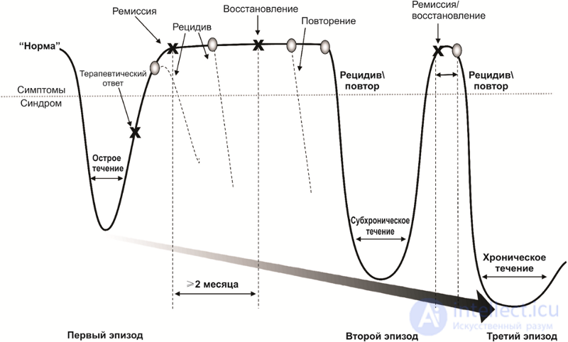 The course of depressive disorders
After a successful course of medication and psychotherapy, a period of remission begins, accompanied either by a weakening (partial remission) or complete disappearance (full remission) of the disease's symptoms. During active treatment and during partial or full remission, there remains a risk of the return of symptoms — relapse.
In addition, there are risks of repeat episodes of depression after full recovery from the first episode. Repeat episodes may be characterized by a different set of symptoms and a more severe course. The disease may become chronic. Often, the first episode of a depressive disorder is psychogenic (reactive), while repeat episodes (if the problem is unresolvable, or the traumatic factor was too intense or prolonged) are endogenous.
Figure 2 outlines the main tenets of the monoamine theory of depression. The formulas of monoamines are given to explain the name of this group of substances — they contain only one amino group (-NH2).
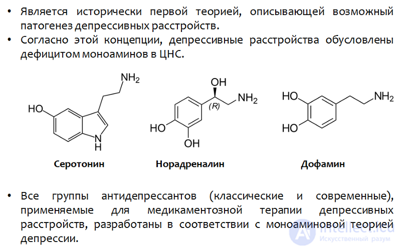 Fig. 2 The monoamine theory of depression
**Serotonin's other NH group is not an amino group; it is part of the indole heterocycle.*
It is believed that the role of monoamines in the formation of individual symptoms of depression is not uniform. For instance, feelings of guilt and worthlessness, suicidal ideation, and appetite disturbance may be attributable to serotonin deficiency.
Dopamine and norepinephrine are responsible for apathy, executive dysfunction, and fatigue.
A deficit of all monoamines together is associated with depressed mood, psychomotor dysfunction, and sleep disturbance.
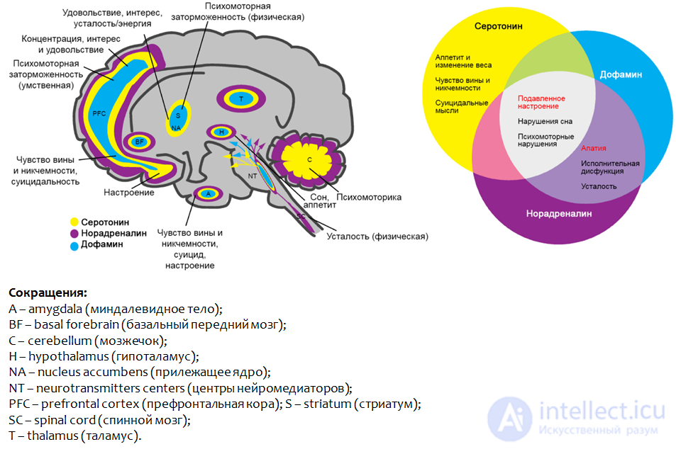 Fig. 3. Saltiel P.F., Silvershein D.I. Major depressive disorder: mechanism-based prescribing for personalized medicine // Neuropsychiatr Dis Treat. 2015. 11. P. 875–88.
Figure 3 shows the brain regions where disruption of the function of the monoamines presented leads to the development of depressive symptoms.
As we have already mentioned, all currently clinically effective antidepressants were developed within the framework of the monoamine theory of depression.
The mechanism of action of antidepressants can loosely be divided into two groups:
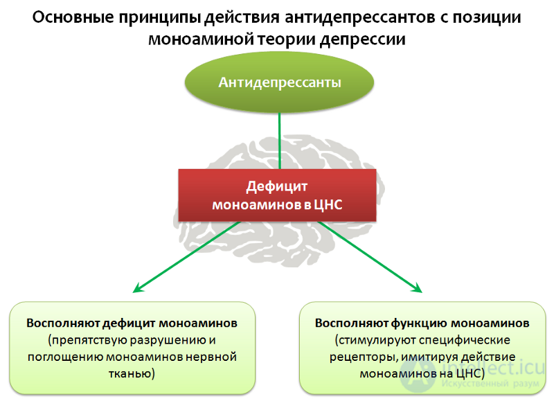 Fig. 4
Let us examine in more detail the main groups of molecular mechanisms of antidepressants. Figure 5 shows the synaptic contact between two nerve cells: at the top — the nerve terminal of one neuron (synapse), at the bottom — another nerve cell that receives the signal.
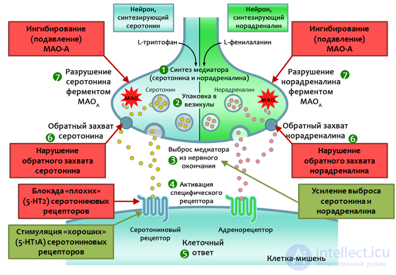 Fig. 5. Drug Therapy of Depression and Anxiety Disorders. Goodman and Gilman’s The Pharmacological Basis of Therapeutics. Twelfth Edition. 2011. Stahl S.M. Basic psychopharmacology of antidepressants. Part 1: Antidepressants have seven distinct mechanisms of action // The Journal of Clinical Psychiatry. 1998. 59. Suppl 4. P. 5–14.
In nerve cells, neurotransmitters (serotonin and noradrenaline) are synthesized, by means of which cells transmit signals to one another. The starting substances for synthesis are the essential amino acids — L-tryptophan and L-phenylalanine. After synthesis, the transmitters are packaged into special granules — vesicles — inside which they travel to the nerve terminals (synapses) and are stored there.
After the cell receives a certain stimulus, the transmitters are released from the nerve terminal (synapse) into the synaptic cleft — the gap between two nerve cells. On the surface of the «receiving» cell there are special protein structures — receptors (in this case, serotonin and adrenergic receptors) — that bind with the transmitter. After binding, the transmitter activates (stimulates) the corresponding receptor, which leads to changes in the metabolic processes inside the cell and accordingly alters its function (enhancing or suppressing it).
After successfully performing its function, 80% of the transmitter is taken back up into the nerve cell, where part of it is broken down by the enzyme monoamine oxidase type A (MAO-A), while part is repackaged into vesicles for reuse. Reuptake of the transmitter allows a significant reduction in the energy expenditure needed to synthesize the transmitter from amino acids.
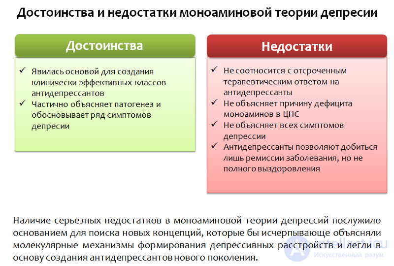 Fig. 6 The drawbacks outweigh the benefits and justify the search for new hypotheses and targets.
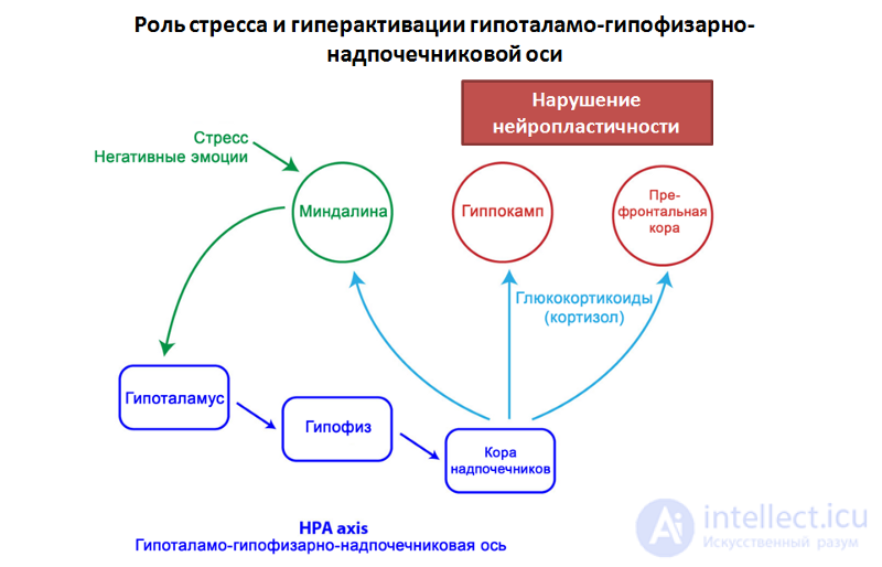
At present, stress is regarded as one of the triggering mechanisms of affective disorders (disorders of the emotional sphere, affect), including depressive disorders. It is believed that what is dangerous is not a single, intense stressful event, but a less intense yet constant exposure to stress, especially everyday unpredictable stressful events. It is impossible to adapt to such stress exposure, and it leads to chronic activation of defense and adaptation mechanisms followed by their exhaustion.
One of the most important components of the body’s physiological response to stress is the hypothalamic–pituitary–adrenal axis (Fig. 7).
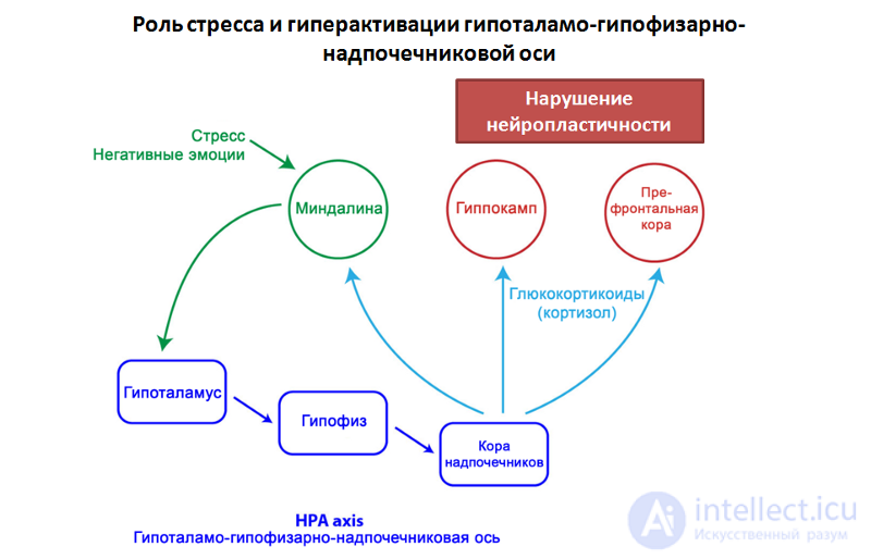 Fig. 7. Varghese F. P. & Brown E. S. The Hypothalamic-Pituitary-Adrenal Axis in Major Depressive Disorder: A Brief Primer for Primary Care Physicians // Primary Care Companion to The Journal of Clinical Psychiatry. 2001. 3(4). P. 151–155.
The sequential stress-induced activation of central structures (the amygdala — the hypothalamus — the pituitary gland) leads to the production of adrenal cortex hormones — glucocorticoids (cortisol) — the stress hormones. These, in turn, are able to act on brain structures responsible for the emotional stress response (the prefrontal cortex and the hippocampus) and disrupt neuroplasticity processes.
Impairments of neuroplasticity lead to disruption of the normal connectivity between brain structures responsible for emotional response.
Neuroplasticity is the brain’s ability to adapt to change through reorganization, both in normal development and under conditions of pathology.
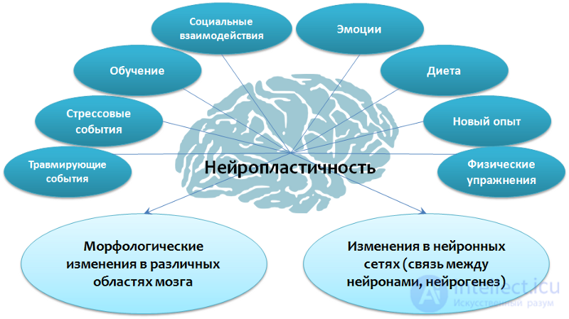 Fig. 8 Fuchs E., Flügge G. Adult Neuroplasticity: More Than 40 Years of Research // Neural Plasticity. 2014. Article ID 541870. Doi:10.1155/2014/541870; Joyce Sh. Neuroplasticity and Clinical Practice: Building Brain Power for Health // Frontiers in Psychology. 7 (2016): 1118. PMC. Web. 7 May 2017. Zilles K. Neuronal plasticity as an adaptive property of the central nervous system // Annals of Anatomy. 1992. Vol. 174. No. 5. P. 383–391.
The prefrontal cortex, the amygdala, and the hippocampus appear to be the most important structures in the context of depressive disorders.
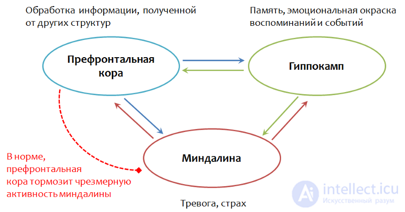 Fig. 9. Gorman J.M., Docherty J.P. A Hypothesized Role for Dendritic Remodeling in the Etiology of Mood and Anxiety Disorders // The Journal of Neuropsychiatry and Clinical Neurosciences. 2010. 22:3. P. 256–264
Normally, when a full-fledged connection exists between the neurons of these structures, the prefrontal cortex processes information received from the hippocampus (memory, the emotional coloring of memories and events). The amygdala is a structure responsible for the sense of fear. Normally, the prefrontal cortex suppresses the excessive activity of this structure.
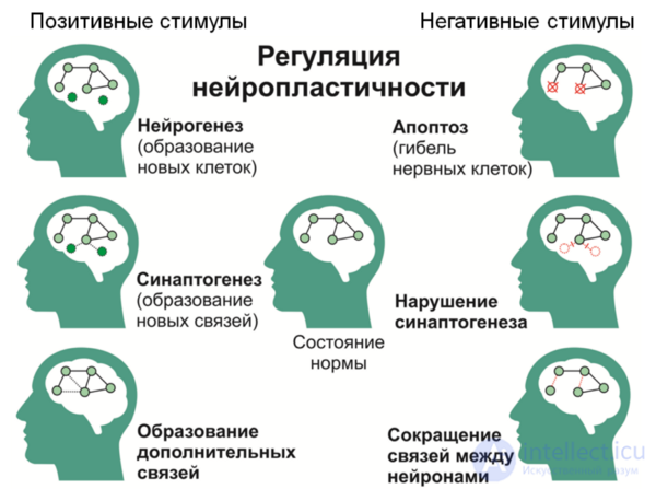 Fig. 10
It is known that against the background of depressive disorders, neuroplasticity processes are disrupted: in particular, the number of contacts between nerve cells decreases, the speed of impulse transmission changes, and the number of neurons decreases. In addition, a decrease in the volume of the hippocampus and prefrontal cortex is observed with depression. Such changes contribute to the disruption of normal functional connectivity between these structures.
Depressive symptoms may apparently be mediated by these changes: uncontrolled anxiety, which is often present in patients with depression, may result from the lack of inhibition of the amygdala by the prefrontal cortex.
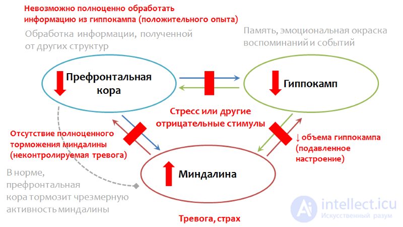 Fig.11. Gorman J.M., Docherty J.P. A Hypothesized Role for Dendritic Remodeling in the Etiology of Mood and Anxiety Disorders. The Journal of Neuropsychiatry and Clinical Neurosciences. 2010. 22(3). P. 256–64. Kudryashov N.V. Experimental study of the psychotropic activity of the pyrazolo[c]pyridine derivative GIZH-72 and the pyrrolodiazepine derivative GMAL-24 under conditions of unpredictable chronic mild stress / Dissertation for the degree of Candidate of Biological Sciences. 14.03.06. Moscow, 2016. 198 p.
The inability to adequately assess a situation and use prior positive experience is a result of disrupted connectivity between the prefrontal cortex and the hippocampus. A decrease in hippocampal volume may explain the pathologically lowered mood.
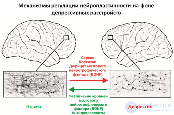 Fig.12
An important regulator of neuroplasticity processes is brain-derived neurotrophic factor (BDNF — brain derived neurotrophic factor), the levels of which decrease under stress and depression.
Stress hormones may also act as negative regulators of neuroplasticity, for example cortisol — a glucocorticosteroid produced by the adrenal cortex. It is well known that most antidepressants in use (with chronic administration) are able to increase BDNF levels, and this apparently forms part of their therapeutic effect.
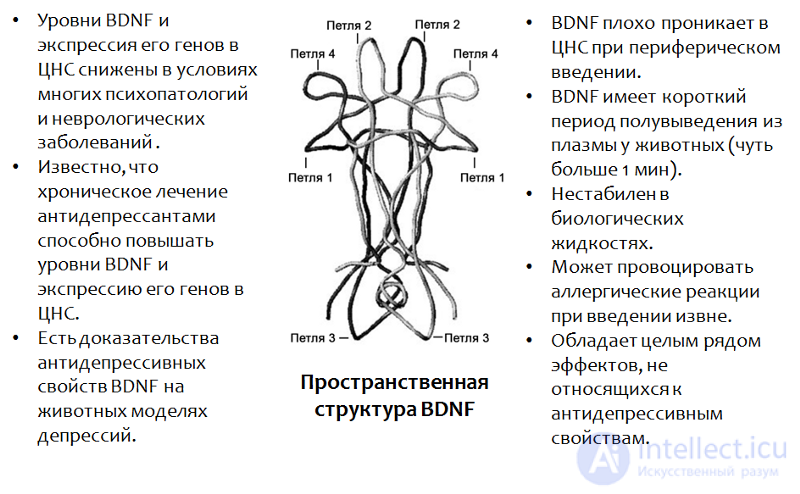 Fig.13. Castrén E., Rantamäki T. The role of BDNF and its receptors in depression and antidepressant drug action: Reactivation of developmental plasticity // Developmental Neurobiology. 2010. 70(5). P. 289–97.
*BDNF plays an important role in many psychopathologies, including depression. The use of BDNF itself is impossible for a number of reasons (which are listed in the figure).
In addition to antidepressants, there are other factors that promote increased BDNF levels in the CNS, and they coincide with positive stimuli for neuroplasticity — learning, physical exercise, new experiences, diet, etc. Moreover, these factors can often complement the pharmacological treatment of depressive disorders.
Figure 14 presents data on a study of the antidepressant properties of BDNF itself in animal models (rats). Since BDNF itself cannot cross into the brain (through the blood-brain barrier) when administered peripherally, in the experiments BDNF was administered directly into the brain.
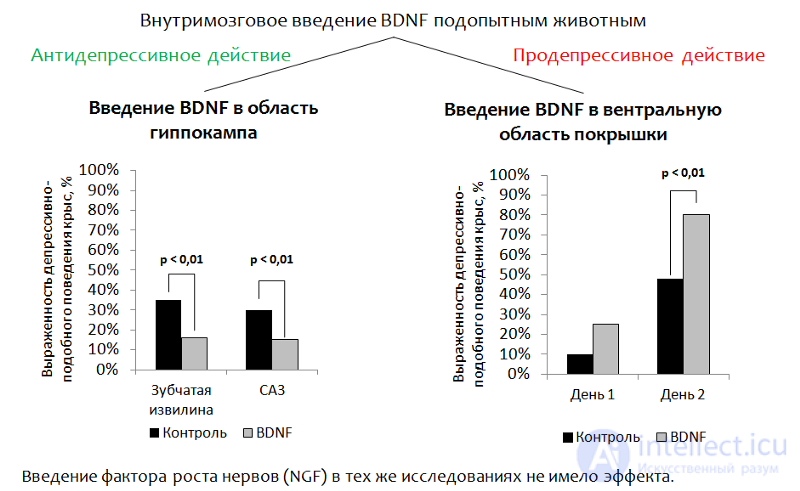 Fig.14 Eisch A.J., Bolaños C.A., de Wit J. et al. Brain-derived neurotrophic factor in the ventral midbrain-nucleus accumbens pathway: a role in depression // Biological Psychiatry. 2003. 54(10). P. 994–1005; Shirayama Y., Chen A.C., Nakagawa S., Russell D.S., Duman R.S. Brain-derived neurotrophic factor produces antidepressant effects in behavioral models of depression. Journal of Neuroscience. 2002. 22(8). P. 3251–61.
1. Administration into the hippocampal region. The main idea was to administer BDNF directly into the region of the brain responsible for neurogenesis (the dentate gyrus of the hippocampus — one of the so-called «neurogenic niches»). After administration, the animals' depressive-like behavior was assessed. *(The dysphoric component of depression is assessed. Animals (rats or mice) are placed in a cylinder of water from which they cannot escape on their own. After some time, the animal's active attempts to escape the cylinder are replaced by a «state of despair» (the animal remains in the water practically motionless). *
Reduced immobility in animals is regarded as a correlate of an antidepressant effect. BDNF produced an antidepressant effect after administration into the dentate gyrus (a neurogenic niche) and the CA3 region of the hippocampus (neurons of this region mediate the interaction of the dentate gyrus with other hippocampal areas).
2. When BDNF was administered into the ventral tegmental area (a region responsible for dopamine production and affected in depressive disorders), the opposite effect was observed — an increase in depressive-like behavior.
Since it is not feasible to use BDNF itself as a medicinal product, drugs based on this factor are being developed. In particular, the active sites of the BDNF molecule have been well studied (their spatial structure gives rise to the name — loop. Fig. 15).
BDNF mimetics (substances that mimic its activity) are currently being actively studied.
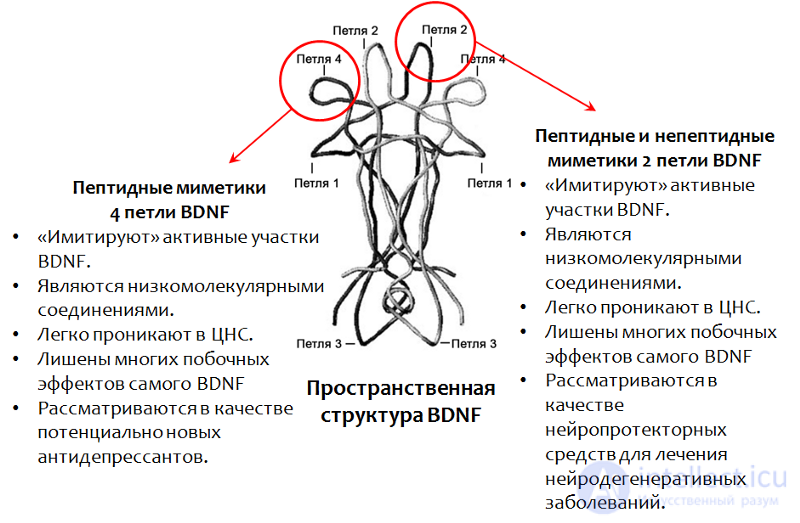 Fig. 15. Fletcher J.M., Morton C.J., Zwar R.A. et al. Design of a conformationally defined and proteolytically stable circular mimetic of brain-derived neurotrophic factor // The Journal of Biological Chemistry. 2008. 283(48). P. 33375–83. Massa S.M., Yang T., Xie Y. et al. Small molecule BDNF mimetics activate TrkB signaling and prevent neuronal degeneration in rodents // The Journal of Clinical Investigation. 2010. 120(5). P. 1774–85. Seredenin S.B., Voronina T.A., Gudasheva T.A. et al. Antidepressant effect of the original low-molecular-weight BDNF mimetic, the dimeric dipeptide GSB-106 // Acta Naturae. 2013. 4(19). P. 116–120.
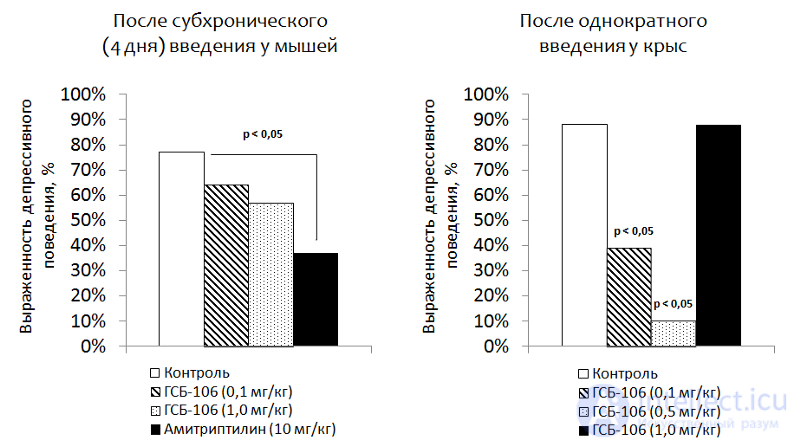 Fig. 16. Seredenin S.B., Voronina T.A., Gudasheva T.A. et al. Antidepressant effect of the original low-molecular-weight BDNF mimetic, the dimeric dipeptide GSB-106 // Acta Naturae. 2013. 4(19). P. 116–120.
GSB-106 is a substance of peptide structure representing a mimetic of loop 4 of BDNF (a domestic Russian development). The substance produces an antidepressant effect in animal models under various administration regimens. Extensive research into the pharmacological properties of this compound is currently underway with the aim of creating a new-generation antidepressant based on it.
*Neurogenesis — a multistage process of forming new nerve cells in the mature CNS, representing an adaptive function of the nervous system.
 Fuchs E., Flügge G. Adult Neuroplasticity: More Than 40 Years of Research // Neural Plasticity. 2014. Article ID 541870, doi:10.1155/2014/541870
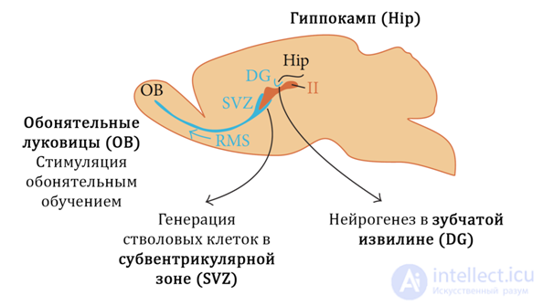 Fig. 17
Figure 17 shows the neurogenic zones (niches) in mammals:
In humans, the hippocampus (dentate gyrus) is considered to be the main neurogenic niche.
Stress, regarded as one of the main triggering stimuli of depressive disorders, leads to decreased BDNF levels and increased cortisol, which in turn enhances the action of glutamate on the CNS.
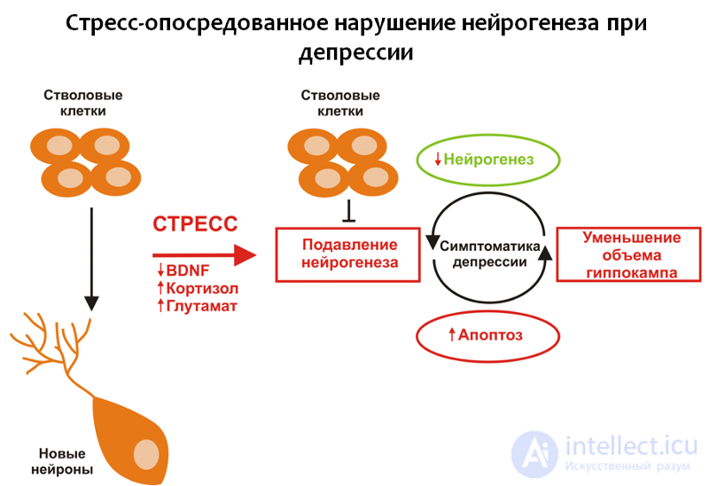 Fig. 18
Taken together, these changes suppress hippocampal neurogenesis and lead to a reduction in hippocampal volume. Glutamate can also activate apoptosis (programmed nerve cell death). When neurogenesis is impaired, the brain cannot fully compensate for these losses, and depressive symptoms develop.
Glutamate is one of the main excitatory amino acids of the CNS. Impairment of neuroplasticity under the influence of excessive glutamate activity appears to be linked to a compensatory response. Neurons «remove» excess connections and die (apoptosis) in order to protect the CNS from overexcitation and the subsequent damaging consequences of this process.
It is a well-known fact that antidepressants can stimulate neurogenesis, although the mechanisms underlying this phenomenon are not yet fully understood. It is known that all classes of antidepressants act on the brain's monoamine system and compensate for functional or material deficits of serotonin and norepinephrine. In addition, drugs of this pharmacological group increase levels of brain-derived neurotrophic factor.
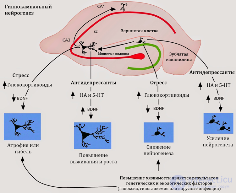 Fig. 19
Stimulation of neurogenesis is characteristic of antidepressants regardless of their mechanism of action, chemical structure, or class. Consequently, the search for mechanisms regulating neurogenesis should focus on properties common to all antidepressants. Such a common property is their activity with respect to serotonin and norepinephrine.
Today, an understanding is forming of the role of serotonin in the regulation of hippocampal neurogenesis.
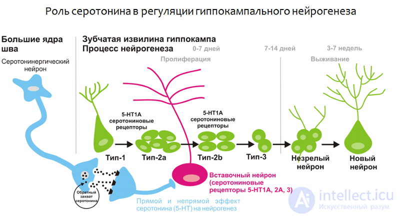 Fig. 20. Alenina N., Klempin F. The role of serotonin in adult hippocampal neurogenesis. Behavioural Brain Research. 2015. 277. P. 49–57.
First, the dentate gyrus of the hippocampus receives serotonergic regulation from the raphe nuclei (a cluster of nerve cells that produce serotonin, located in the brainstem), both directly and via interneurons that carry various subtypes of serotonin receptors on their surface.
Second, serotonin receptors of the 1A subtype have been found on the stem cells themselves, which suggests the potential for serotonin to regulate brain stem cells.
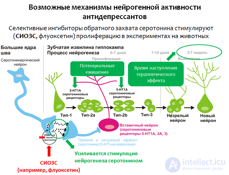 Fig. 21
As has been shown in experimental studies (in animals and cell cultures), selective serotonin reuptake inhibitors (SSRIs, the classic drug of this group being fluoxetine) are able to stimulate the proliferation stage of neurogenesis in the hippocampus.
The proposed mechanism is an increase in the concentration of serotonin (5-HT, i.e., 5-hydroxytryptamine) in the CNS and the subsequent (enhanced) stimulation of neurogenesis by serotonin.
Potential targets of antidepressants in the process of neurogenesis may also include serotonin receptors of the 1A subtype (5-HT1A receptors). These assumptions are consistent with data on the positive (therapeutic) effect of activating serotonin 5-HT1A receptors by antidepressants (for example, vilazodone) in depressive disorders.
Another argument that allows stimulation of neurogenesis to be regarded as the main mechanism of action of antidepressants is the temporal coincidence between the average time of onset of the therapeutic effect (from 2 to 7 weeks) and the full cycle of neurogenesis (3–7 weeks).
In addition to the mechanisms listed, SSRI antidepressants also demonstrate the ability to increase BDNF levels, although the mechanisms of this effect remain unknown.
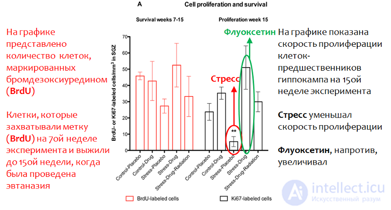 Fig. 22. Perera T.D., Dwork A.J., Keegan K.A., et al. Necessity of Hippocampal Neurogenesis for the Therapeutic Action of Antidepressants in Adult Nonhuman Primates // PLoS ONE. 2011. 6(4):e17600. doi:10.1371/journal.pone.0017600.
Studies in primates (the most relevant animal model) have shown that fluoxetine (trade name "Prozac") is able to stimulate neurogenesis in the context of stress (in this case, a social isolation stress model was used). Figure 22 shows that fluoxetine significantly increased (statistically significantly) the proliferation (division) rate of neural stem cells in the primate hippocampus.
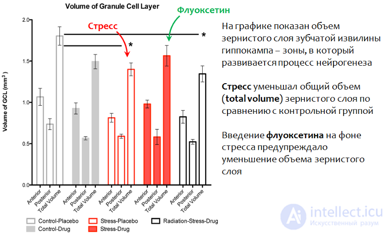 Fig. 23. Perera T.D., Dwork A.J., Keegan K.A., et al. Necessity of Hippocampal Neurogenesis for the Therapeutic Action of Antidepressants in Adult Nonhuman Primates // PLoS ONE. 2011. 6(4):e17600. doi:10.1371/journal.pone.0017600.
Exposure to stress led to a decrease in the granule cell layer of the hippocampal dentate gyrus — the main neurogenic zone of the primate brain. Administration of fluoxetine during stress exposure prevented this change and preserved the normal volume of this structure (total volume).
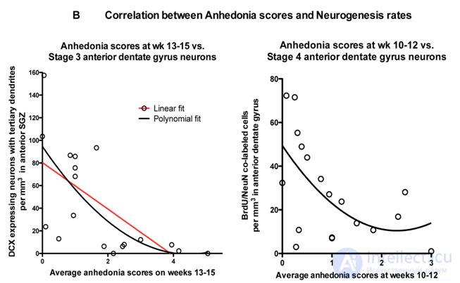 Perera T.D., Dwork A.J., Keegan K.A., et al. Necessity of Hippocampal Neurogenesis for the Therapeutic Action of Antidepressants in Adult Nonhuman Primates. PLoS ONE. 2011. 6(4):e17600. doi:10.1371/journal.pone.0017600.
A correlation was established between the stimulation of neurogenesis by fluoxetine and the prevention of the development of depressive-like behavior (anhedonia).
In studying the effect of fluoxetine on neurogenesis in nonhuman primates, a significant correlation was established between decreased neurogenesis (caused by stress) and increased depressive-like behavior in primates (anhedonia, which was determined by a combination of disturbances in social and feeding behavior), as well as a correlation between the stimulation of neurogenesis by fluoxetine and the absence of depressive-like behavior.
Norepinephrine is another important monoamine that, along with serotonin, can participate in the regulation of hippocampal neurogenesis.
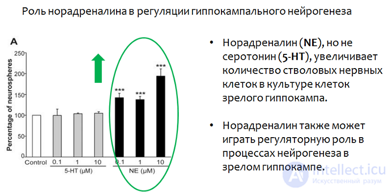 Fig. 25. Jhaveri D.J., Mackay E.W., Hamlin A.S., et al. Norepinephrine directly activates adult hippocampal precursors via β3 adrenergic receptors // The Journal of Neuroscience. 2010. 30(7). P. 2795–2806. doi:10.1523/JNEUROSCI.3780–09.2010.
Studies on hippocampal neuron cultures have shown that norepinephrine, unlike serotonin, increased the number of stem cells. Serotonin, as was demonstrated earlier, affected not the number, but the rate of proliferation.
In addition to quantitative changes, norepinephrine also caused qualitative ones — it increased the size of neurospheres, as clearly shown in the electron microscope image (see fig. 26).
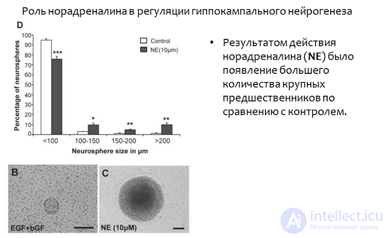 Fig. 26. Jhaveri D.J., Mackay E.W., Hamlin A.S., et al. Norepinephrine directly activates adult hippocampal precursors via β3 adrenergic receptors // The Journal of Neuroscience. 2010. 30(7). P. 2795–2806. doi:10.1523/JNEUROSCI.3780–09.2010.
Earlier we examined changes in neurons, but overlooked the role of glial cells in the formation of depressive disorders. Nevertheless, experimental and clinical studies point to a possible role of glial cell pathology in the pathogenesis of depression.
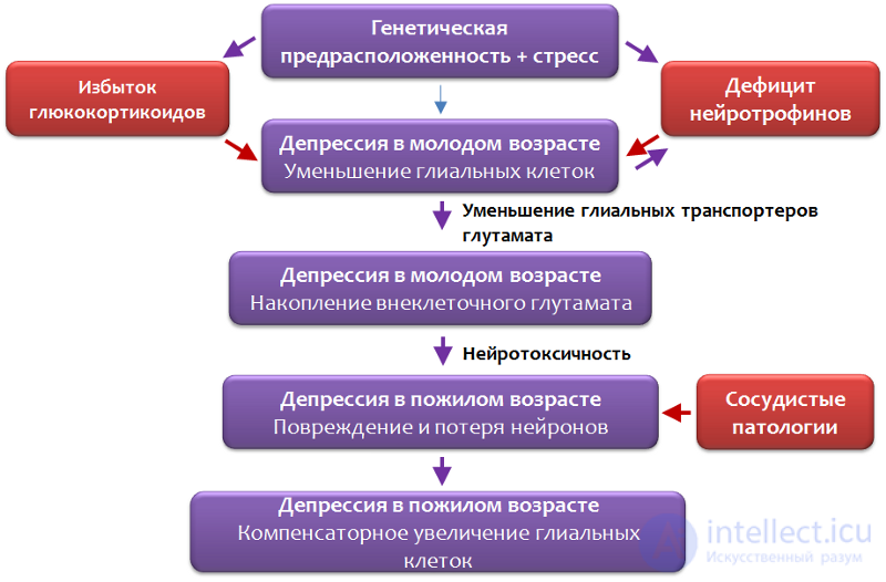 Fig. 27. Rajkowska G., Miguel-Hidalgo J. J. Gliogenesis and Glial Pathology in Depression // CNS & Neurological Disorders Drug Targets. 2007. 6(3). P. 219–233.
Researchers have proposed a scheme for the pathogenesis of depressive disorders involving glia (Fig. 27).
Genetic predisposition may include: a hereditary factor (having a parent with a depressive disorder increases the child's risk of the disease); polymorphisms of genes such as BDNF, the serotonin transporter (which is involved in the reuptake of serotonin into neurons and glial cells), serotonin receptors, and enzymes of serotonin synthesis (tryptophan hydroxylase type 2).
Genetic vulnerability, together with environmental and stress factors, creates a favorable environment for the development of a depressive disorder.
Scientists have established that the role of glial cells differs between young and elderly patients (Fig. 28). Glial cells may play an important role in the pathogenesis of the early stages of depressive disorders, which may account for the pronounced decrease in the number of pyramidal neurons at a more mature age.
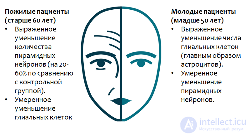 Fig. 28. Rajkowska G., Miguel-Hidalgo J. J. Gliogenesis and Glial Pathology in Depression // CNS & Neurological Disorders Drug Targets. 2007. 6(3). P. 219–233.
In particular, loss of glial cell function at a young age increases the risk of recurrent depressive episodes in old age, but in that case a deficit of pyramidal neurons, rather than glial cells, will predominate.
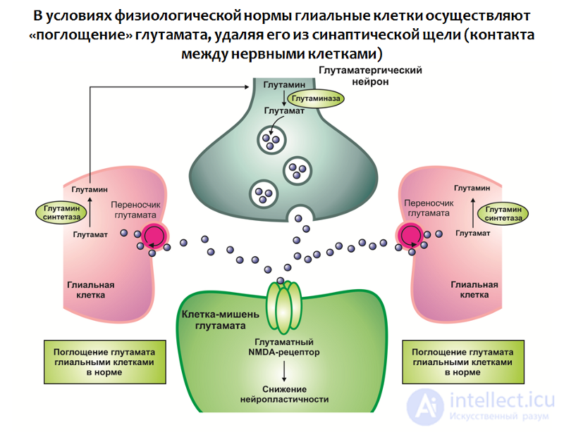 Fig. 29. Rajkowska G., Miguel-Hidalgo J. J. Gliogenesis and Glial Pathology in Depression // CNS & Neurological Disorders Drug Targets. 2007. 6(3). P. 219–233.
This dynamic is consistent with one of the most important functions of glial cells — absorbing excess glutamate from the synaptic cleft (the contact between nerve cells). Glutamate is one of the main excitatory neurotransmitters of the CNS, and its excessive action can lead to impaired neuroplasticity and excitotoxicity (neurotoxicity associated with excessive excitation; it appears to be a protective reaction of nerve cells against overexcitation — the number of neurons and the connections between them decreases).
Glial cells contain a transporter protein that participates in carrying glutamate from the synaptic cleft into the glial cell, where glutamate undergoes metabolism.
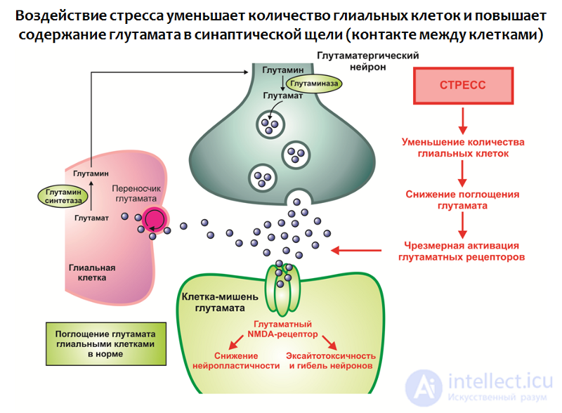 Fig. 30. Rajkowska G., Miguel-Hidalgo J. J. Gliogenesis and Glial Pathology in Depression // CNS & Neurological Disorders Drug Targets. 2007. 6(3). P. 219–233.
Exposure to stress, together with other factors, leads to:
An excess of this excitatory neurotransmitter is thus formed, which acts as a negative modulator of neuroplasticity (it is believed that this may be part of a compensatory reaction protecting the nervous system from overexcitation).
The function of glial cells is not limited to glutamate uptake; they are also involved in producing neurotrophins, in particular BDNF (Fig. 31).
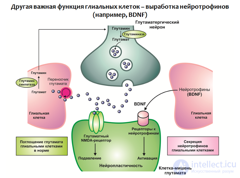 Fig. 31
Together, a deficit of neurotrophins and an excess of glutamate lead to impaired neuroplasticity and depression-like changes (a reduction in the volume of the hippocampus and prefrontal cortex, and disruption of normal functional connectivity between structures of the limbic circuit).
Within the framework of this concept, it is also possible to find explanations for the therapeutic efficacy of antidepressants (Fig. 32):
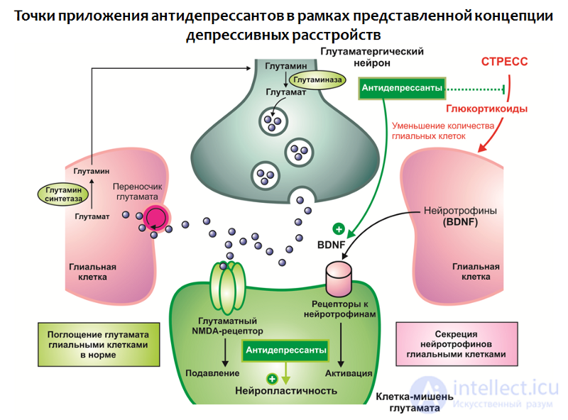 Fig. 32
Figure 33 presents a generalized scheme of depressive disorders, built on the concept of stress-mediated neurodegeneration. It can be seen that antidepressants occupy the niche of «correctors of the consequences of stress». Despite all their merits and therapeutic potential, antidepressants are not always effective in eliminating depressive symptoms.
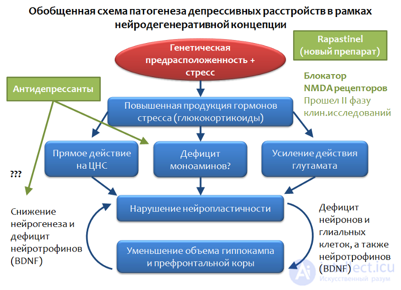 Fig. 33
There exist so-called treatment-resistant forms of depressive disorders. This phenomenon has been explained by the diversity of stress factors, by the varying strength and duration of stress exposure, and by individual characteristics (mutation of antidepressant targets). But the overall conclusion from this situation is the same — a search for fundamentally new targets for the pharmacotherapy of depressive disorders is needed.
A promising direction appears to be action on the glutamate system, if this neurotransmitter is considered one of the key elements in the pathogenesis of depressive disorders. Significant progress has been achieved within this direction — a fundamentally new antidepressant has been created that, in terms of its mechanism, is a blocker of glutamate NMDA receptors and prevents excessive activity of this amino acid. The antidepressant Rapastinel has now successfully completed phase I and II clinical trials, where it demonstrated high efficacy and is regarded as a treatment for resistant forms of depressive disorders.
Within the framework of the glutamatergic theory of depressive disorders, one can also consider the role of the main inhibitory mediator of the CNS — gamma-aminobutyric acid (GABA).
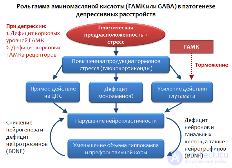 Fig. 34. Möhler H. The GABA system in anxiety and depression and its therapeutic potential // Neuropharmacology. 2012. Jan. 62(1). P. 42–53.
GABA is the functional opposite of glutamate and is able to limit its excitatory action, so assessing the role of GABA in depressive disorders seems entirely logical.
It has been established that against the background of depressive disorders, a deficit of cortical GABA levels and its receptors is observed. In particular, pyramidal neurons that produce glutamate can be subject to inhibitory influence from interneurons that produce GABA. GABA exerts its inhibitory action through activation of the GABA-A receptor.
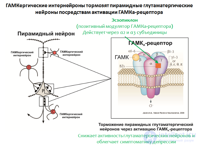 Fig. 35. Möhler H. The GABA system in anxiety and depression and its therapeutic potential // Neuropharmacology. 2012. Jan. 62(1). P. 42–53.
The structure of the GABA-A receptor is shown in Figure 35. The receptor consists of 5 subunits (2α, 2β and γ), each subunit having a subtype; for example, 6 variants of the α-subunit are known. The combination of different subunit variants determines the subtype of the GABA-A receptor.
The role of GABA is also supported by the efficacy of a positive modulator of the GABA-A receptor — eszopiclone. The target of this drug is GABA-A receptors that contain α2– and α3-subunits.
Eszopiclone
is sometimes used in combination with antidepressants and significantly alleviates depressive symptoms even after discontinuation of antidepressants. It is thought that its therapeutic action is related to attenuation of glutamate function. Interestingly, other positive
продолжение следует...
Часть 1 Depression: Causes, Diagnosis, Prevention and Treatment; Depression in Neurology and Neurosurgery, and During Pregnancy, Childbirth,
Часть 2 Depression during pregnancy, childbirth, and menopause - Depression: Causes, Diagnosis,
Часть 3 The «ideal» drug - Depression: Causes, Diagnosis, Prevention and Treatment;

Comments