
Lecture
Objective: to build students' knowledge of the main types of wounds and haemorrhages and their characteristics, the main stages of first aid for wounds and haemorrhages, and to build skills in providing first aid; to foster in students a sense of responsibility and moral readiness for unexpected and extreme situations.
Wounds. A wound is damage to tissues in which the integrity of the skin, mucous membranes, or deeper-lying tissues is disrupted. That is, disruption of the integrity of the skin, mucous membranes, deeper-lying tissues and the surface of internal organs as a result of mechanical or other action is called a wound, or an open injury.
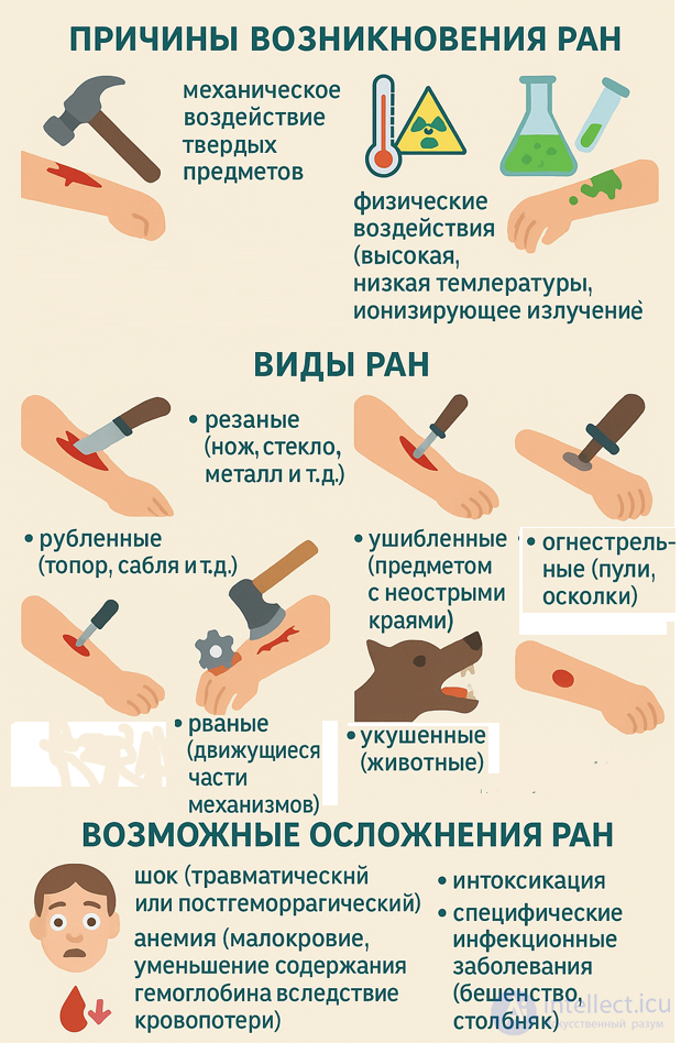
Causes of wounds:
- mechanical action of hard objects;
- physical action (high or low temperature, ionising radiation);
- chemical action (acid, alkali).
Types of wounds:
- incised (knife, glass, metal, etc.);
- chopped (axe, sabre, etc.);
- stab (awl, nail, bayonet, etc.);
- bruised (an object with blunt edges);
- lacerated (moving parts of mechanisms);
- bite (animals);
- gunshot (bullets, fragments).
Possible complications of wounds:
- shock (traumatic or post-haemorrhagic);
- anaemia (reduced blood, decreased haemoglobin content due to blood loss);
- intoxication;
- specific infectious diseases (rabies, tetanus).
SUPERFICIAL AND DEEP WOUNDS ARE DISTINGUISHED. Deep wounds in which the internal linings of cavities (abdominal, thoracic, cranial, joint) are damaged are called PENETRATING. All other types of wounds, regardless of their depth, are called NON-PENETRATING. All wounds, except wounds inflicted by a sterile instrument during surgery, should be considered INFECTED. Depending on the nature of the wounding object, WOUNDS ARE DISTINGUISHED AS STAB, BITE, INCISED, CHOPPED, BRUISED, LACERATED, AND GUNSHOT WOUNDS. Every wound is characterised by pain and bleeding.
Haemorrhage is the outflow of blood from the bloodstream into the external environment or internal organs. Normally, a person has about 4-5 litres of blood, of which 60% circulates through the vessels and 40% is held in the blood depot (liver, spleen, and others). Loss of 1/3 of the blood is life-threatening, but patients can also die from a smaller blood loss if it occurs rapidly. Men tolerate blood loss worse, while women are better adapted to blood loss.
Depending on the type of damaged blood vessels, bleeding can be: arterial, venous, capillary, parenchymal (internal), or mixed.
ARTERIAL BLEEDING - bleeding from damaged arteries. THE BLOOD FLOWING OUT IS BRIGHT RED IN COLOUR AND IS EJECTED IN A STRONG PULSATING STREAM. Arterial bleeding from a small artery can be successfully stopped with a pressure dressing. FOR THE EMERGENCY STOPPING OF ARTERIAL BLEEDING, DIGITAL COMPRESSION OF THE ARTERY IS WIDELY USED. (a - carotid; b - submandibular; c - temporal; d - subclavian; e - brachial; f - axillary). The artery can be pressed with the thumb, palm, or fist. PRESSING ARTERIES BY FIXING THE LIMB IN A CERTAIN POSITION is used during transportation of the patient to hospital. Bleeding from arteries is reliably stopped by tight circular compression of the limb, ensuring compression of all vessels above the site of injury. This is most easily accomplished with the help of a special rubber tourniquet.
VENOUS BLEEDING occurs when veins are damaged. The pressure in veins is significantly lower than in arteries, so blood flows out slowly, in an even or uneven stream. IN THIS TYPE OF BLEEDING, THE BLOOD IS DARK CHERRY-COLOURED. In venous bleeding, reliable temporary stopping of the bleeding is achieved by applying a pressure dressing.
CAPILLARY BLEEDING occurs when the smallest blood vessels - capillaries - are damaged. Capillary bleeding is easily stopped by applying an ordinary dressing to the wound.
EXTERNAL AND INTERNAL BLEEDING ARE DISTINGUISHED.
EXTERNAL BLEEDING is characterised by blood entering directly onto the surface of the body through a wound in the skin.
In INTERNAL BLEEDING, blood enters some cavity: the free abdominal cavity; haemorrhages into internal organs (intraorganic haematomas); gastrointestinal bleeding.
Internal bleeding is a cause for urgent hospitalisation. If internal bleeding is suspected, an ambulance should be called as quickly as possible, and first aid should be given before it arrives. Possible internal bleeding can be identified by the following signs: pallor of the skin; fatigue, drowsiness, weakness; coughing with clots of blood or scarlet frothy sputum in pulmonary haemorrhage; "coffee-ground" vomiting or dark tarry stool in gastric bleeding; cold sweat; decreased blood pressure and increased pulse rate.
Haemorrhage is the outflow of blood beyond the bloodstream into tissues, some body cavity, or the external environment.
|
Type of bleeding |
Brief characteristic |
|
arterial |
scarlet-coloured blood, flows out in a pulsating stream |
|
venous |
dark-coloured blood, flows out in a steady stream |
|
capillary |
the entire wound surface bleeds, blood flows out in small drops (like dew) |
|
parenchymal |
the entire wound surface of the parenchymal organ (liver, spleen, lung, kidney, etc.) bleeds |
The circulating blood volume (CBV) is 5-8% of body weight. For example: if body weight is 70 kg, then the CBV is 3.5-5 L. Approximately 80% of the CBV circulates through the vessels. 20% of the CBV is held in the vascular depot (liver, spleen, skin).
General disorders of vital activity occur with acute blood loss of more than 10% of the CBV, and so-called post-haemorrhagic shock develops.
The general signs of acute blood loss are:
- pale skin;
- cold sweat;
- rapid pulse;
- low blood pressure;
- dry mouth, thirst;
- rapid breathing;
- increasing weakness, nausea.
Methods of temporarily stopping bleeding
|
mechanical |
elevated position of the limb (for capillary, venous) |
|
digital compression of the damaged vessel (for arterial) |
|
|
pressure dressing (for venous) |
|
|
maximum flexion of the limb (for arterial) |
|
|
tourniquet, twist tourniquet (for arterial) |
|
|
physical |
cold applied to the site of bleeding (for venous, capillary, parenchymal) |
|
dusting with a sorbent (for venous, capillary) |
Sequence of first-aid measures for wounds with external bleeding:
The algorithm for providing first aid for internal bleeding should be approximately as follows:
By the amount of blood loss in any bleeding: Mild blood loss; Moderate blood loss; Severe blood loss; Extremely severe blood loss
Arterial bleeding from the vessels of the upper and lower limbs is stopped in two stages:
First, the artery above the site of injury is pressed against the bone (closer in the direction of the heart) to stop blood flow to the wound site, and then a standard tourniquet or one made from improvised materials is applied. The following should not be used as a tourniquet: electrical wires, cords and other narrow materials, which can lead to damage to soft tissues, blood vessels, and nerve trunks.
Arteries are best pressed against bony prominences at specific points where the pulse is easily felt.
The temporal artery is pressed with the thumb at the temple, in front of and slightly above the ear.
The carotid artery is pressed on the left or right (only on one side!) on the lateral surface of the neck.
The subclavian artery is pressed in the fossa above the collarbone against the first rib.
The axillary artery (for bleeding from a wound in the area of the shoulder joint and forearm) is pressed against the head of the humerus along the anterior hairline in the armpit.
The brachial artery is pressed against the humerus on the inner side of the biceps.
The radial artery is pressed against the underlying bone in the wrist area near the thumb.
The femoral artery is pressed in the area of the inguinal fold, at its midpoint.
The popliteal artery is pressed in the area of the popliteal fossa.
The arteries of the dorsum of the foot are pressed against the underlying bone.
After pressing the artery, application of the haemostatic tourniquet begins. The tourniquet is applied over clothing or over fabric placed underneath it. Applying the tourniquet to bare skin is not permitted. The tourniquet is placed on the limb above the site of bleeding, approximately 3-5 cm from the wound, stretched strongly and, without reducing the tension, tightened around the limb and its ends secured. When the tourniquet is applied correctly, bleeding from the wound stops, the limb below the site of application turns pale, and the pulse in the artery disappears. A note indicating the date, hour and minute of its application must be placed under the tourniquet.
The limb below the site of tourniquet application retains its viability for 2 hours, and in winter outdoors for 1-1.5 hours, so after this time has elapsed the tourniquet must be removed and, after a few minutes, reapplied, but slightly above the previous site.
Possible errors in applying a tourniquet:
- too weak a tightening causes compression of the veins only, as a result of which arterial bleeding intensifies;
- too strong a tightening, especially on the upper arm, leads to damage to nerve trunks and paralysis of the limb;
- applying the tourniquet directly to the skin usually leads, after 40-60 minutes, to severe pain at the site of its application.
A casualty with acute blood loss is pale, covered in cold sweat, usually lethargic, indifferent to their surroundings, speaks in a quiet voice, complains of dizziness and "spots" flickering or darkening before the eyes when raising the head, asks for water, and notes dryness in the mouth. The pulse is rapid, of poor fullness, blood pressure is lowered, and with rapid loss of a large amount of blood, a picture of haemorrhagic shock with persistent hypotension develops.
Shock most often occurs as a result of severe, extensive injuries accompanied by blood loss. Predisposing factors for the development of traumatic shock are nervous and physical overexertion, cooling, and radiation injuries. Traumatic shock can occur with injuries not accompanied by significant bleeding, especially if the most sensitive, so-called reflexogenic zones are injured (thoracic cavity, cranium, abdominal cavity, perineum).
Signs: the picture of traumatic shock consists of two phases.
The first - excitation is often not noticed. This is a fleeting motor reaction of the casualty (while conscious). The face is pale, the gaze restless, there is speech agitation - complaints, shouting, breathing is rapid and uneven, the pulse is tense.
The second phase - mental depression, indifference to the surroundings, a sharp decrease in reaction to pain while consciousness is preserved, the pulse is rapid and weak, temperature is lowered, breathing is rapid and shallow, the skin is covered with sticky, cold sweat. The onset of vomiting is a sign of a particularly dangerous condition of the casualty.
THE SECOND PHASE OF SHOCK IS DIVIDED INTO FOUR DEGREES:
SHOCK OF DEGREE I (MILD). The casualty is pale, consciousness is usually clear, sometimes with mild lethargy, reflexes are reduced, and there is shortness of breath. PULSE IS RAPID, 90-100 BEATS PER MINUTE.
SHOCK OF DEGREE II (MODERATE). Pronounced lethargy, sluggishness. PULSE 120-140 BEATS PER MINUTE.
SHOCK OF DEGREE III (SEVERE). Consciousness is preserved, but the surroundings are not perceived. The skin is earthy-grey in colour and covered with cold, sticky sweat, and cyanosis of the lips, nose and fingertips is pronounced. PULSE 140-160 BEATS PER MINUTE.
SHOCK OF DEGREE IV (PRE-AGONAL OR AGONAL STATE). Consciousness is absent. Pulse cannot be detected.
First aid: it is necessary to reduce pain as quickly as possible, stop the bleeding, and immobilise the fracture. To reduce pain, it is recommended to use ethyl chloride, analgin (2-3 tablets), cardiac agents - ephedrine, cordiamine, valerian solution. If breathing worsens - artificial respiration (mouth to mouth). Warming the body. Hot sweet tea or water should be given. 100-120 ml of 40% alcohol (vodka) may be given.
The next most important task of first aid is organising the swiftest transportation of the casualty to hospital. It is best to transport in a special resuscitation vehicle, in which effective measures can be carried out.
It should be remembered that shock is easier to prevent than to treat, so when providing first aid to an injured person it is necessary to carry out
Treatment tactics must be differentiated and depend on the specific type of bleeding. There is a general set of measures that must be carried out for any type. All specific manipulations are targeted in nature, since performing them incorrectly can harm the patient.
General measures of aid for bleeding include:
The cause of most deaths after injury is acute blood loss, THEREFORE THE FIRST MEASURES MUST BE AIMED AT STOPPING THE BLEEDING BY ANY POSSIBLE MEANS (pressing the vessel, a pressure dressing, and in cases of pronounced arterial or venous bleeding - applying a tourniquet, etc.). An equally important task of FIRST AID is PROTECTING THE WOUND FROM CONTAMINATION AND INFECTION. Proper wound treatment prevents the development of complications in the wound and reduces its healing time by almost 3 times.
WOUND TREATMENT SHOULD BE CARRIED OUT WITH CLEAN, PREFERABLY DISINFECTED HANDS. WHEN APPLYING AN ASEPTIC DRESSING, THE LAYERS OF GAUZE THAT WILL COME INTO DIRECT CONTACT WITH THE WOUND SHOULD NOT BE TOUCHED WITH THE HANDS. The wound can be protected simply by applying an aseptic dressing (bandage, individual dressing packet, triangular bandage). The skin around the wound is treated with 5% iodine tincture, and any loose foreign bodies are removed from the wound.
When providing first aid to a patient with a penetrating chest wound, communication between the pleural cavity and the external environment must be stopped as early as possible with the help of an occlusive dressing.
More than a hundred years ago, the French scientist Pasteur proved that infection of wounds occurs as a result of the entry into wounds of the smallest living organisms, visible only under a microscope - disease-causing microbes, or, as is often said, the entry of infection. Microbes are found in the air and on all objects surrounding a person. A person is constantly in contact with a huge number of microbes. The most diverse microorganisms can be found on the skin and mucous membranes. However, they penetrate into the body only when the integrity of the skin or mucous membranes is disrupted as a result of wounds, abrasions, punctures, burns, or a decrease in the protective properties of the skin and mucous membranes due to impaired blood supply, cooling, exhaustion, and weakening of the body due to general illnesses. Microbes that have penetrated into the tissues of the body cause local purulent-inflammatory phenomena at the site of entry (wound suppuration, abscesses, phlegmons), and in more severe cases, when infection breaks through into the blood - general infection (sepsis). Suppuration is most often caused by microbes shaped like spheres (staphylococci, streptococci) and rods (intestinal, pyocyanic, and others). Most surgical manipulations (operations, injections, blockades, intravenous and subcutaneous infusions, etc.) are accompanied by some disruption of the integrity of the skin, as a result of which the penetration of infection into the body becomes possible.
Prevention of wound infection and combating infection that has entered a wound are carried out with the help of a complex of measures known as "antisepsis" and "asepsis".
Asepsis - a method of combating surgical infection, the basis of which is the prevention of infection entering the wound. This is achieved by the complete decontamination of all objects that may come into contact with the wound.
The destruction of microbes and their spores on surgical operating linen, instruments, suture and dressing materials, gloves, gowns and the hands of surgeons is called sterilisation. Sterilisation is carried out by various methods: steam under pressure - autoclaving, dry heat, calcination, boiling, flaming, and soaking in antiseptic solutions and antibiotic solutions.
Sterilisation by radioactive radiation (gamma rays), ultraviolet rays (mercury-quartz lamps), gases, etc. is also fairly widely used.
An object is considered sterile if there are no microbes capable of reproducing on its surface or throughout its thickness. The sterility of objects is monitored by means of bacteriological cultures on special nutrient media.
Antisepsis - a complex of measures aimed at destroying microbes in a wound and creating conditions in the wound that are unfavourable for the development of microbes and their penetration deep into the tissues.
Antisepsis is carried out by mechanical, physical, chemical and biological methods. Mechanical antisepsis consists of removing dead and crushed tissues, blood clots, and foreign bodies from the wound. One example of mechanical antisepsis is primary surgical treatment of the wound, which is performed by a doctor in a surgical hospital. Physical antisepsis: quartz irradiation of wounds, insertion into the wound of various drains, swabs and gauze strips moistened with hypertonic sodium chloride solutions, ensuring the outflow of pus and wound fluid outward into the dressing, which creates unfavourable conditions for the development of infection in the wound. This method of antisepsis is also mainly used in medical care.
Chemical and biological antisepsis are of the greatest importance in providing first aid, i.e. the use of various substances that cause the destruction of microbes that have entered the wound or slow down their reproduction (bactericidal substances).

Comments