
Lecture
LASER THERAPY
Laser therapy is the therapeutic use of low-intensity laser radiation in the ultraviolet, visible, and infrared spectrum.
Laser radiation in an optically transparent medium is characterized by monochromaticity (a strictly defined wavelength), coherence (the radiation phase is constant in time and space), high directionality (a very small beam divergence angle), and polarization (a fixed orientation of the electromagnetic field vectors in space).
The main components of a laser are the pump (excitation) source of the working substance, the active medium capable of transitioning into an excited (induced) state (the working substance), a resonator that allows the laser radiation to be repeatedly amplified and concentrated, and a power supply unit.
Atoms of the working substance, absorbing electromagnetic energy from the pump source, transition into an excited state in which they cannot remain for long. The avalanche-like process of the working substance's atoms transitioning from the excited to the unexcited state is accompanied by the emission of laser radiation of the same frequency, phase, and direction as the inducing radiation.
Depending on the working substance used, lasers are divided into solid-state, liquid, gas, and semiconductor types. Laser radiation can be obtained in the infrared, visible, and ultraviolet regions of the spectrum.
The main factors causing the biological tissue's reaction to laser radiation are monochromaticity and the associated high spectral power density (all the radiation energy is concentrated in a very narrow frequency range). Such specific properties of laser radiation as polarization and coherence do not play a significant role in the mechanism of the laser's therapeutic action, since laser radiation loses them already in the superficial layers of biological tissue, which is not an optically transparent medium.
In clinical practice, lasers of the red and infrared radiation spectra have found the main application. Red-spectrum lasers (0.63-0.69 um) are used to act on mucous membranes, skin, and adjacent tissues. For exposure to deep-lying tissues and organs, the infrared laser radiation range of 0.8-0.95 um is universal. In reflexotherapy, lasers of the red (0.63-0.69 um) and infrared (1.2-1.3 um) spectra are used.
The mechanism of the therapeutic action of low-energy laser radiation has not yet been fully studied. It is assumed that the action of laser radiation is based on the interaction of light with photosensitizers (acceptor molecules) in body tissues. These molecules are able to selectively absorb light quanta of a certain wavelength, resulting in the excitation of biomolecule electrons and their transition to a singlet state. From this state they can pass to the ground state with the emission of a light quantum (fluorescence), or to a long-lived triplet state, which differs from the singlet state in its reactivity, allowing biomolecular complexes to actively participate in various cellular metabolic processes. The transition of biomolecules from the triplet state to the ground state is accompanied by the emission of a light quantum, i.e., so-called re-emission occurs.
In animal and human tissues, the photoacceptors of red radiation are DNA molecules (absorption maximum at a wavelength of 0.620 um), cytochrome oxidase (0.600 um), cytochrome (0.632 um), superoxide dismutase (0.630 um), and catalase (0.628 um). Radiation of the near-infrared range is absorbed predominantly by nucleic acid molecules (0.820 um). Red and infrared radiation are also absorbed by oxygen.
The transfer of laser excitation energy to biomolecules can also occur through non-radiative exchange between electronically excited molecules (photodonors) and molecules in the ground state (photoacceptors), by the so-called inductive-resonance and exchange-resonance methods. g
The interaction of laser radiation with biological molecules is most often realized at cell membranes, leading to a nonspecific reaction of the cells of the irradiated tissue: a change in the surface charge of cells and their dielectric permeability, an increase in the activity of enzymatic and metabolic processes, an increase in the level of tissue oxygen consumption and the oxidation-reduction potential, and an enhancement of bioenergetic and biosynthetic processes.
Activation of these processes stimulates the synthesis of proteins and nucleic acids, glycolysis, lipolysis, and oxidative phosphorylation of cells. Activation of plastic processes and accumulation of macrophages leads to an increase in oxygen consumption and enhanced intracellular oxidation of organic substances, i.e., an improvement of trophism in the irradiated tissues. Due to vasodilation, local blood flow is normalized, which leads to dehydration of the inflammatory focus, stimulates reparative processes in tissues, and increases the adhesive activity of neutrophils.
As a result of conformational changes in the proteins of the voltage-gated ion channels of the neurolemma of cutaneous afferents, laser radiation causes suppression of tactile sensitivity. A decrease in the impulse activity of the nerve endings of C-afferents from the pain focus leads to a reduction in pain sensitivity. In addition to local reactions, afferent impulses from cutaneous and muscular nerve receptors form, through segmental-metameric connections, reactions of internal organs and surrounding tissues, as well as generalized reactions of the whole organism (activation of endocrine glands, cellular and humoral immunity, and reparative processes).
When laser irradiation of circulating blood occurs, activation of the enzyme systems of erythrocytes takes place, leading to an increase in the oxygen capacity of the blood. Action on the nuclei and membranes of cells stimulates the differentiation and functional activity of irradiated blood cell elements. The rate of platelet aggregation decreases, the anticoagulant system is suppressed, which causes a significant slowdown in the rate of thrombus formation and improves blood microcirculation. Clinically, laser irradiation has a clearly pronounced stimulating, desensitizing, anti-inflammatory, decongestant, and analgesic effect.
Indications:
— diseases of the cardiovascular system (coronary heart disease, hypertension, occlusive arterial diseases of atherosclerotic origin, thrombophlebitis);
— diseases of the bronchopulmonary system (bronchial asthma, chronic bronchitis);
— diseases of the digestive system (chronic cholecystitis, chronic hepatitis with moderate impairment of liver function, chronic pancreatitis, peptic ulcer of the stomach and duodenum);
— diseases of the nervous system (neuralgia, neuritis, traumatic injuries of peripheral nerves, ganglionitis);
— diseases of the musculoskeletal system (rheumatoid arthritis, osteoarthrosis, spinal osteochondrosis, traumatic injuries);
— diseases of the urogenital system (chronic prostatitis, cervical erosion, chronic salpingo-oophoritis);
— skin diseases (trophic ulcers of various origins, long-term non-healing wounds, burns in the healing stage);
— diseases of the ear, throat, and nose (chronic rhinitis, chronic pharyngitis, chronic laryngitis, chronic tonsillitis, chronic maxillary sinusitis, otitis);
— diseases of the oral mucosa (periodontal disease, gingivitis);
— eye diseases (keratitis, corneal ulcer);
— thymus-dependent immunodeficiency states.
Contraindications: malignant neoplasms, blood diseases, thyrotoxicosis, infectious diseases, cachexia, bleeding, acute and subacute stages of myocardial infarction and cerebral circulatory disorders, circulatory insufficiency of stage IIB-III, crisis course of hypertension, disseminated tuberculosis, functional renal insufficiency, liver cirrhosis.
Equipment, general instructions for performing procedures
At present, a large number of laser physiotherapy devices are manufactured. Only lasers of the red and infrared spectra have found practical application.
Red-spectrum radiation is generated by helium-neon lasers «UFL-01», «Yagoda», «AFL», «LYUZAR», «FALM», «ADEPT», «Alok-1» and «Alok-2» (wavelength 0.63 um), as well as semiconductor lasers «Azor-2K» and «Mustang» (wavelength 0.65-0.68 um). All red-spectrum lasers have low output power values (2-30 mW) and generate continuous or modulated-intermittent radiation, with the power in the pulse not increasing.
Infrared-spectrum radiation (wavelength 0.8-0.95 um and 1.2-1.3 um) is generated in continuous mode by semiconductor lasers «Kolokolchik», «Mlada», «Izel», «Mustang», «Azor-2K», and in pulsed mode by «Uzor-2K», «MILTA», «Lita-1», «ELAT», «Mustang», «Azor-2K». With a low average output power (5-10 mW), the pulse power of these lasers reaches values of 6-12 W or more. The pulse repetition rate for most devices is 1-3000 Hz.
Many laser devices are universal, since they generate red and infrared radiation spectra in both continuous and pulsed modes («Mustang», «Azor-2K», «Skalyar», «ADEPT»). They are also equipped with ring-shaped magnetic attachments that create a magnetic field with a magnetic induction of 20-30 mT at the surface of the attachment.
Devices of other radiation spectra (nitrogen, argon, helium-cadmium, copper-vapor) are currently represented by experimental models. Clinical experience with their use is still insufficient. Such devices include the laser physiotherapy device «ATL-1» based on solid-state dyes, which generates laser radiation in the frequency spectrum from yellow to infrared (wavelength 0.55-1.05 um) with a pulse duration of 25 ms, a pulse repetition rate of 1-3 Hz, and a radiation energy of 0.5 mJ per pulse. In terms of the effectiveness of its analgesic and anti-inflammatory action, it surpasses all other industrially manufactured laser devices of the visible and infrared radiation spectra. -
Dosing. The general principles of therapeutic exposure to physical factors apply to laser radiation. Small exposure doses produce a stimulating effect, medium doses produce an analgesic effect and improve microcirculation, and increased doses produce an anti-inflammatory, inhibitory effect. Large doses have a negative effect, leading to exacerbation of the pathological process.
The exposure dose with continuous red-spectrum laser devices is determined by the power flux density, expressed in mW/cm2- and by the exposure time in minutes, or by the energy flux density, expressed in J/cm2(recall that energy is defined as the product of power and time).
The power flux density of laser radiation is measured with special measuring instruments «IM-1» and «IM-2». During measurements, care must be taken to ensure that the laser radiation falls on the irradiated surface strictly perpendicularly, otherwise part of the radiation will be reflected from the surface.
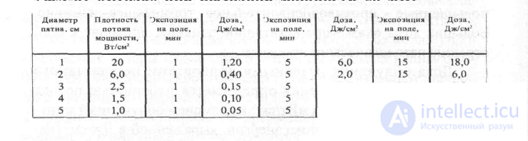
At the same output power of a laser device, the power flux density varies depending on whether the laser beam is focused or defocused, which leads to a change in the activity and depth of the therapeutic effects. At a light spot diameter of 1 cm, the power flux density reaches 20 mW/cm2and more. In this case, the optimal exposure time is 3-5 min. When the light spot diameter is 5 cm, the power flux density does not exceed 1 mW/cm2, and the exposure time should be increased to 10-15 min per treatment zone.
Clinical observations have established that at an exposure dose of 0.5 J/cm2, stimulation of reparative processes in damaged tissues is most clearly manifested. At an exposure dose of 1-2 J/cm2an analgesic, sedative effect appears, and microcirculation is stimulated. At an exposure dose of 2-3 J/cm2an anti-inflammatory effect is pronounced. A dose of 4 J/cm2and more per field (pathological focus) is not recommended for use. Approximate exposure doses with a helium-neon laser are given in Table 4.
Table 4
Exposure dose with the helium-neon laser device UFL-01 «Yagoda» at an output power of 20 mW
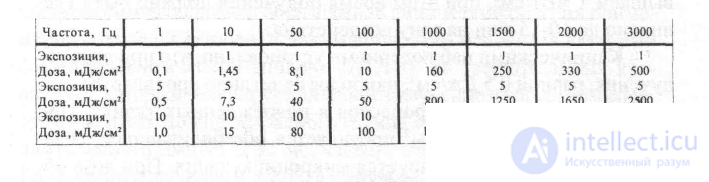
More complex is the dosing of pulsed infrared radiation, whose therapeutic effects depend not only on the average power and exposure time, but also on the pulse power and pulse repetition rate. Low pulse repetition rates (from 1 to 50 Hz) have a stimulating effect on reparative processes and cell function (glands of internal and external secretion, etc.); frequencies of 100-150 Hz produce an analgesic and stimulating effect; frequencies of 1000-3000 Hz have a pronounced anti-inflammatory effect. Laser radiation with a frequency above 1000 Hz is apparently perceived by biological tissue as continuous. Approximate exposure doses with pulsed infrared lasers are given in Table 5.
Table 5
Exposure dose with the infrared pulsed laser device "Lita-1"
In invasive irradiation of circulating blood, the exposure dose can be 3.5 J/cm2, for which, at an output power of 2 mW at the end of the light guide, intravenous irradiation is carried out for 30 min, r:
Procedure technique. Radiation from helium-neon lasers is delivered to the irradiated object using a mirror reflector-diffuser or a light guide, while radiation from semiconductor infrared lasers is delivered using light guides, remote emitting heads, and various optical attachments that are mounted on open emitting heads. Irradiation is performed remotely or by contact, using either a stable or a labile (scanning) technique.
The skin is exposed to a helium-neon laser remotely, from a distance of 10-50 cm, with the power flux density regulated by the diameter of the light spot. The same area (zone) can be irradiated using a stable or labile exposure technique. In the stable technique, the area to be irradiated is divided into sections, and the laser beam, after the prescribed exposure time, is moved from one section to the adjacent one, with the exposure time repeated. In the labile technique, the laser beam is moved over the exposure zone throughout the entire procedure, either manually or by a scanning device.
When the skin is exposed to a semiconductor laser, the stable technique is used on acupuncture points or small joints; in other cases, the labile technique is used. As a rule, exposure is carried out by the contact method, with the emitting head moved slowly in circular motions over the entire zone to be irradiated. Remote exposure is carried out using scanning devices.
Irradiation of the mucous membranes of the nasal cavity, paranasal sinuses, oral cavity, vagina, and rectum is carried out using light guides and various optical attachments.
For irradiation of the gastric and duodenal mucosa, the epithelium of the trachea and bronchi (endoscopic method), and circulating blood (invasive method), flexible fiber-optic light guides are used.
For the laser therapy procedure, the patient is placed on a couch or seated on a chair, depending on the location of the irradiation site. The distance from the optical attachment to the irradiated body surface with the remote technique is from 10 to 50 cm. With IR lasers, the technique is contact-based. The irradiated surface must be bare, cleared of ointment and cream. Exposure to infrared laser radiation on wound surfaces through thin gauze dressings (0.2-0.5 cm) is permitted. The laser beam must fall perpendicularly on the irradiated surface.
Warning: given that the main property of laser radiation is its powerful stimulating action, laser therapy should be used with extreme caution in cases of suspected malignant neoplasms, as well as in the postoperative period after removal of a malignant tumor.
Safety precautions. During the laser therapy procedure, the eyes of medical staff and the patient must be protected from direct and reflected laser radiation by goggles made of blue-green SZS-22 glass (GOST 9411-81E).
Some procedure techniques
Exposure to the cardiac region
The method is indicated for coronary heart disease and neurocirculatory dystonia of the cardiac type.
Helium-neon laser radiation is applied remotely, using the stable technique. Exposure fields: the anterior surface of the chest wall in the region of the cardiac apex, the interscapular region to the left of the spine, the middle of the left sternocleidomastoid muscle, and the inner surface of the left shoulder. The diameter of the laser beam spot is 5 cm (power flux density 1 mW/cm2). The duration of exposure of each zone is 1-3 min. The exposure time is increased every 3 procedures. The course of treatment is 12-15 procedures.
Infrared laser radiation is applied by contact, using the stable technique (laser puncture). Points (zones) of exposure: the middle of the left sternocleidomastoid muscle, the second intercostal space to the right and left of the sternum, the fourth intercostal space along the left midclavicular line, and three points paravertebrally on the left at the level of ThIII-ThIV. The exposure mode is pulsed, frequency 5-50 Hz, pulse power 4-6 W, exposure time 1 min per zone. The course of treatment is 10-12 procedures.
Exposure to the joints
With helium-neon laser radiation, large joints are irradiated from three sides and small joints from two sides. The exposure technique is stable and remote. For irradiation of large joints, the diameter of the laser beam spot is 5 cm (power flux density 1 mW/cm2), exposure duration 8-10 min. 1-2 joints may be irradiated per day. For irradiation of small joints, the diameter of the laser beam spot is 2-3 cm (power flux density 10 mW/cm2), exposure duration 1-2 min per joint. 2-3 joints are irradiated per procedure. The course of treatment is 12-15 procedures.
Infrared laser radiation on the joints is prescribed by contact, using the scanning technique. Continuous laser radiation or pulsed radiation with a frequency of 1500 Hz and a pulse power of 6-10 W is used. Exposure time is 4-6 min for a large joint and 2-3 min for a small one. 1-2 large joints or 3-4 small joints are irradiated per procedure. The course of treatment is 10-12 procedures
Exposure to slow-healing wounds, trophic ulcers The wound (ulcer) is cleared of ointment, purulent discharge, and necrotic plaque. Irradiation of the wound (ulcer) surface with a helium-neon laser is carried out remotely, using the stable technique. The operating mode of the laser device is continuous or pulsed with a frequency of 10 Hz. The diameter of the laser beam spot is 7-5 cm (power flux density 0.5-1 mW/cm2), exposure time 5-8 min. The wound edges are then irradiated using the labile technique for 1-2 min at a power flux density of 5-10 mW/cm2. Procedures are carried out daily. The course of treatment is 20-30 procedures.
Irradiation of the wound (ulcer) surface with infrared laser is performed remotely, using a labile technique. The gap between the emitting head and the wound (ulcer) surface is set equal to 1 cm, and the wound surface may be covered with a sterile gauze pad. The irradiation mode is pulsed, with a frequency of 10-80 Hz and a pulse power of 10 W, exposure 10 min. Course of treatment 15-20 procedures.
Supra-arterial irradiation of circulating blood.
In psychoneurological diseases, the author uses the method of supra-arterial irradiation of blood with red and infrared laser radiation.
Helium-neon radiation or radiation of the red spectrum is applied remotely. Zone of exposure: the anterior surface of the neck in the area of projection of the carotid arteries. The diameter of the laser beam spot is 2 cm, radiation intensity up to 10 W/cm2. Duration of exposure is 10 min for each carotid artery.
+When exposed to infrared radiation, the emitters are placed in contact with the projection zone of the carotid artery. The irradiation mode is continuous («MLT», «Mlada»,» NUR») or pulsed at a frequency of 1,5-3000 Hz. Duration of exposure is 10 minutes on each carotid artery daily. Course of treatment 10-12 procedures.
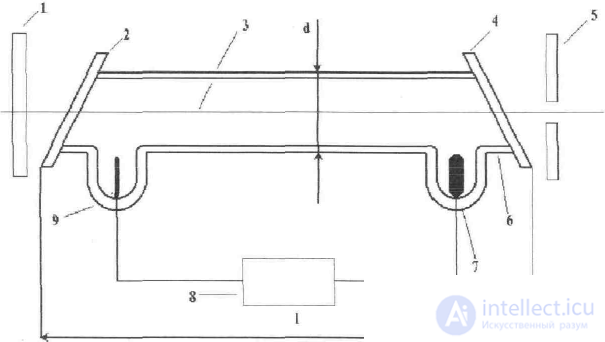
Gas (helium-neon) laser consists of three fundamentally important units: the emitter, the pumping system and the power supply, whose operation is ensured with the help of special auxiliary devices. A simplified structural diagram of a helium-neon laser is shown in the figure below.
The emitter is designed to convert pump energy (transferring helium-neon mixture 3 into the active state) into laser radiation and contains an optical resonator, which in the general case is a system of carefully manufactured reflecting, refracting and focusing elements, in whose internal space a certain type of electromagnetic oscillations of the optical range is excited and maintained. The optical resonator must have minimal losses in the working part of the spectrum and high manufacturing precision of the components and their mutual alignment. In the laser shown in the figure, the optical resonator is made in the form of two parallel mirrors 1 and 5, located outside the active part of the medium 3, which is separated from the surrounding environment by the bulb 6 of the discharge tube and two windows 2, 4 with plane-parallel boundaries forming Brewster's angle with the radiation axis. The external mirrors 1 and 5 provide multiple passage of the radiation through the active medium with increasing power of the laser radiation flux. For the radiation to exit, one of the mirrors (5) is made with a hole or semi-transparent.
The pumping system is designed to convert the energy of the electric power source 8 into the energy of the laser's ionized active medium 3. Pumping is carried out by an electric discharge, for which two electrodes are installed in it - cathode 7 and anode 9, between which voltage is applied from the power source. Helium atoms are excited upon collisions with fast electrons and, colliding with neon atoms, transfer their energy to them. In some types of lasers, focusing magnets or coils and special diversion tubes for circulating the active medium are used.
A typical representative of mass-produced laser (solid-state)physiotherapy devices is the «Orion».
Orion"-a unique laser device created specifically for home treatment. It combines high treatment efficiency with ease of use, reliability and absolute safety. "Orion" is thirty years of scientific research and clinical trials embodied, with the help of modern electronic technologies, in a small and elegant device. The “Orion” laser has laser safety class 1.
Infrared laser treatment allows laser blood irradiation sessions to be performed without breaching the integrity of the skin. List of indications for use:
Surgical pathologies, injuries;
Eczema, herpes, psoriasis, pyodermatitis, pustular skin diseases, urticaria;
Acne, rhinophyma, cellulite, seborrhea, scars;
Stomatitis, gingivitis, pulpitis, periodontitis, periodontal disease;
Sore throat (angina), tonsillitis, pharyngitis, laryngitis, sinusitis, otitis;
Tracheitis, bronchitis, pneumonia, bronchial asthma;
CHD (coronary heart disease), myocarditis, angina pectoris, arrhythmia, hypertension;
Gastritis, ulcer, esophagitis, colitis, pancreatitis, cholecystitis;
Hemorrhoids, proctitis, cystitis, urethritis, prostatitis, adenoma;
Gynecological diseases, menopausal syndromes, ovarian hypofunction;
Sexual disorders;
Obliterating endarteritis, diabetic angiopathy, atherosclerosis, thrombophlebitis, varicose veins;
Arthritis, arthrosis, osteochondrosis, heel spur, myositis;
Neuroses, vegetative-vascular dystonia, migraine, neuralgia, neuritis;
Allergic manifestations;
Rejuvenating and health-improving procedures.
Treatment with "Orion" will become even more effective and comfortable thanks to special attachments, created by Russian scientists in collaboration with doctors. The attachments enhance the directionality of the radiation and shape the desired "form" of the laser field. This expands the boundaries of laser therapy application and promotes faster recovery.
|
|
||||||||||
|
|
|
||||||||||
|
|
||||||||||
|
|
At present, the device «Matrix» is manufactured, allowing the use of laser attachments, magnetic, EHF
Parameters of emitting heads of various types:
|
|
|
|
|||||||||||||||||||||||||||||||||||||||||||||||||||||||||||||||||||||||||||||||||||||||||||||||||||||||||||||||||||||||||||
|
|
LED emitting heads:
Emitting heads of the EHF range:
|
|
|||||||||||||||||||||||||||||||||||||||||||||||||||||||||||||||||||||||||||||||||||||||||||||||||||||||||||||||||||||||||||
|
|
|
|
The choice of emitting heads is made in accordance with the required spectral range of radiation and power. Thus, for exposure to acupuncture points, heads with an emitting power of more than 8 mW are not used.
Optical and magnetic attachments for laser therapy of the device «Matrix»
Emitting heads with a single laser source make it possible to use optical attachments to introduce the laser radiation directly into the light channel without the use of special optics, by means of a simple threaded or collet connection (a «rigid» instrument). At the output of the attachments, the required distribution of the light flux delivered to the desired location is obtained.
For laser therapy, a set of instruments is sufficient, containing the following radiation patterns: «cone», «wide cone», «side cone», «sphere», «cylinder». The required scattering diagrams, depending on the task, are formed by shaping the distal end of the light guide into a specific geometric form, or by introducing a metal reflector into the cavity of the protective sheath of the distal end.
The light-guide instrument consists of three main parts: a mounting connector, a rod, and the working part — an optical scattering element. From the optical connector to the diffuser, the radiation travels through the light guide. The diffuser ensures convenient fixation within the cavity of the pathological focus and its uniform irradiation.
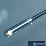
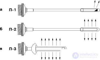
Proctological attachment P-1 (fig. 1, a)forms a radiation spot Ø 5—10 mm at an angle of 120°, which makes it possible to obtain a local power density distribution. Used for irradiation of the prostate gland (rectally).
Proctological attachment P-2 (fig. 1, b)distributes the radiation evenly over a cylinder Ø 9 mm and 25 mm long. Used in urology for irradiation of the prostate gland (rectally) or in proctology for irradiation of the rectal walls. Due to scattering over a large surface, the power density is significantly lower than that of attachment P-2, which is compensated to some extent by its versatility.
Proctological attachment P-3 (fig. 1, c)distributes the radiation evenly over a cylinder Ø 9 mm and 25 mm long. Used in proctology for irradiation of the rectal walls (anal fissures, hemorrhoids, etc.).
Fig. 1. Proctological attachments
|
|
|
|
|
|
|
|
|
|
|
|
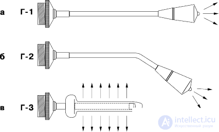
Gynecological attachments G-1 (fig. 2, a) and G-2 (fig. 2, b), used for intravaginal irradiation of the cervix and adnexa, scatter radiation over an area Ø 15—20 mm in contact. Gynecological attachment G-3 (fig. 2, c) is used intravaginally for certain inflammatory diseases.
Fig. 2. Gynecological attachments
|
|
|
|
|
|
|
|
|
|
|
|
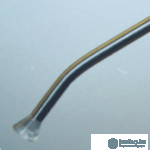
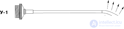
Urological attachment U-1 (fig. 3) is designed for transurethral exposure of the prostate gland and urethra. Made of flexible material, the attachment length is 30 cm. The scattering cylindrical region at the end is 20 mm long.
Fig. 3. Urological attachment U-1 for LO-type heads
|
|
|
|
|
|
|
|
|
|
|
|
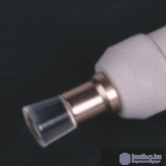
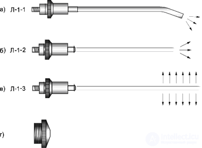
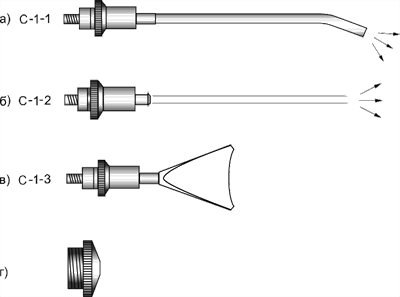
Attachments L-1 — ENT (fig. 4) and S-1 — dental (fig. 5) are supplied as kits.
Fig. 4. Set of ENT attachments: a — L-1-1, b — L-1-2, c — L-1-3, d — adapter device
|
|
|
|
|
|
|
|
|
|
|
|
|
|
|
|
|
|
Fig. 5. Set of dental attachments: a — S-1-1, b — S-1-2, c — S-1-3, d — adapter device |
|
|
|
|
|
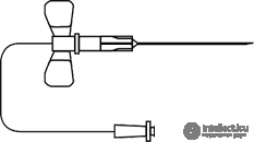
The disposable sterile light guide KIVL-01 (fig. 6) is a length of polymer fiber, one end of which is glued into a plastic cylinder that provides self-alignment of the light guide, while the other end is inserted into a needle for intravenous injections. This device allows the procedure of intravenous laser blood irradiation to be carried out as quickly and effectively as possible [Pat. 2252048 RU]
Fig. 6. Disposable light guide with needle for ILBI
|
|
|
|
|
|
|
|
+Fig. 7. Acupuncture attachment A-3
|
|
|
|
|
|
|
|
Physiological basis of the therapeutic use of the devices .the pathogenetic conditionality of the effect is provided by anti-inflammatory, analgesic, anti-edema, desensitizing, immunocorrective effects, improvement of the rheological properties of blood and vascular microcirculation.
For skin diseases, external diseases of a purulent nature, and impaired tissue epithelialization in wound and burn injuries, toilet of the affected surface is performed beforehand. The technique is remote-stable, with a pulse repetition rate – 1500 Hz. Irradiation is carried out in fields – around the periphery of the pathological focus, capturing healthy tissues. Exposure time is up to 3 minutes per field. Total irradiation time per procedure is up to 20 minutes. Procedures are performed daily or, according to indications, twice a day, especially in the initial period of the disease. Course of treatment:10-15 procedures.
For diseases of the musculoskeletal system and peripheral nervous system, irradiation is performed by contact, stably, or by scanning. Exposure per field is 3-7min. The number of irradiation fields is up to 6. procedures are performed daily or, according to instructions, twice a day. In the acute period, twice a day with minimal exposures. Course of treatment: 15-20 procedures.
For diseases of the internal organs (chronic gastritis, gastric and duodenal ulcer, prostatitis, ischemic heart disease, chronic lung diseases, etc.), a technique of irradiating reflexogenic zones and the projection area of the organ is used. The technique is contact-stable, with a pulse repetition rate of 1500 Hz. Irradiation time per field is 2-4 min. Total irradiation time per procedure is up to 15 min. Course of treatment: 10-15 procedures.
For inflammatory diseases ( otitis, rhinitis, pharyngitis, tonsillitis) the pathological focus itself is irradiated using optical attachments, or the projection area. The technique is contact-stable. Irradiation time per zone is 2-4 min. Total irradiation time per procedure is up to 10min. Procedures are performed daily. In the acute period, it is advisable to perform procedures twice a day with exposures of 1-2 min. Course of treatment: 10-15 procedures.
List the contraindications to the use of the low-frequency laser «Orion»?
By what criteria are the power, exposure, and method of irradiating the patient selected?
Describe the sequence for preparing the device for operation?
What is the maximum continuous operating time of the device?
How should irradiation of the musculoskeletal system be performed?
For what purposes is the magnetic attachment used?
What is the advantage of the contact-stable irradiation method over others?






















Comments