
Lecture
To understand the effect of electric current on the body, it is necessary to know the properties of tissue.
Electrical properties of various tissues are not the same. The liquid media of the body - cerebrospinal fluid, blood, intercellular fluid - are characterized by electrical conductivity with respect to direct and low-frequency alternating current. Nerves conduct current well along nerve fibers. Bone without periosteum has the greatest resistance to electric current. The epidermis of the skin, connective tissue formations, ligaments, and tendons have high resistance to current. These tissues can rightfully be classified as insulators or dielectrics. The nature of such a conductor is resistive-capacitive. At a frequency of 2kHz the resistance is 796 Ohm, while at a frequency of 5kHz it is only 32 Ohm. Hence the conclusion that as frequency increases above 3-4 Hz, skin resistance becomes insignificant for it. The same can be said regarding cell membranes and tendons. These areas are good conductors.
Body tissues practically do not absorb the energy of a constant magnetic field. An alternating magnetic field induces eddy currents in tissues with good electrical conductivity, which generate a certain amount of heat per unit time. At ν=50Hz this heat is extremely small compared to the heat generated as a result of tissue metabolism.
The impossibility of separating the specific and nonspecific effects of physical factors, as well as their ability to produce several physiological effects at once, some of which are common to a number of factors, makes it difficult to classify them according to their physiological effect. As a result, physical factors are classified by type of energy and the nature of the physical action. In particular, in electrotherapy, methods are distinguished based on the use of electric currents and electric, magnetic, and electromagnetic fields.
Galvanization is the therapeutic use of a constant electric current of low voltage (up to 80 V), which does not change in magnitude, at a low current strength 9 to 50 mA).
The method came into use shortly after the invention, in 1800, of a source of direct current by the Italian physicist A. Volta, who called the current galvanic. The therapeutic method was likewise named after the current. Nowadays, galvanization uses exclusively current obtained by rectifying and smoothing alternating mains current. The amplitude of the resulting ripple must not exceed 0.5%.
Due to the high ohmic resistance of the stratum corneum of the epidermis, current enters the body mainly through the openings of the sweat glands and, to a lesser extent, the sebaceous glands; and since their total area is only about 1/200 of the skin's surface, most of the energy of the applied current is expended in passing through the epidermis. It is here that the most pronounced primary reactions to current exposure also develop.
Having overcome the resistance of the epidermis and subcutaneous fatty tissue at the sub-electrode areas of the body, the current, in the form of oppositely directed movement of oppositely charged ions moving from electrode to electrode, passes through the tissues with the least resistance, branching considerably and deviating from a straight line between the two electrodes. It is hardly possible to predict in advance which paths it will take — whether in parallel bundles [Shchedrakov V. I., 1959; R. Peterson, 1966], along blood vessels, along nerves, or through muscle tissue [Orlov A. N., 1977, et al.] Apparently, in each individual case these will be different paths depending on the area of exposure, the variants of the individual's anatomical structure, and the functional state of the body's systems.
Along the current's path at semipermeable membranes, including cell membranes, an accumulation of like-charged ions occurs on both sides of them. Between such accumulations of ions of opposite polarity, an intratissue polarization current of reverse direction arises. On the one hand, this creates additional resistance to the acting current, and on the other — such areas within the tissues are the sites of the most active (after the epidermis) action of the current.
Mechanisms of the therapeutic effect. Encountering the high resistance of the epidermis, the energy of the direct current is partly converted into heat and partly causes primary electrochemical processes. Because the current intensity is low, the amount of heat generated as it passes through the skin is insignificant. However, it can produce weak biological effects in the form of activated blood circulation and enhanced biochemical processes. The main and specific component of the action of direct current, however, is its effect on the ratio of various ions in the tissues, which is one of the important links in the regulation of their functional state. This action of direct current is fully explained from the standpoint of the ionic theory of excitation developed by V. Yu. Chagovets (1957), P. P. Lazarev (1923), and A. N. Obrosov (1955, 195H). This theory, in turn, is based on the theory of electrolytic dissociation developed in 1883—1887 by the Swedish physical chemist and natural scientist S. Arrhenius, according to which in electrolyte solutions there is a constant breakdown of neutral molecules into positively and negatively charged particles — ions — and, in parallel, their recombination into neutral molecules. Building on this theory, J.Loeb, as a result of experimental studies, established that for the normal state of various tissues, as well as for their excitation, the decisive factor is not so much the concentration as the quantitative ratio between mono- and divalent ions, in particular between the amount of sodium and potassium ions, on the one hand, and calcium and magnesium ions – on the other:
[Na]+ + [K]+/ [Ca]+ + [Mg]+
When this ratio increases due to a rise in the amount of potassium and sodium ions, excitation occurs. When this ratio decreases due to an increase in calcium and magnesium ions, the intensity of vital processes in the tissues decreases.
When electrodes are placed on the head, depending on their location, reactions characteristic of excitation of the gustatory (sensation of a metallic taste) or visual (appearance of phosphenes) analyzers may occur. Reactions of the vestibular apparatus in the form of dizziness and unsteadiness are also possible. Experimental studies of head galvanization have revealed excitation of cortical cells of the cerebrum and synaptic activity of individual neurons [Vorontsov D.S., 1958]. The use of various exposure intensities has shown that at a current density of 0.5 mA/cm2 gross circulatory disturbances occur in the brain. At an intensity of up to 0.1 mA/cm2 stimulation of protective-compensatory mechanisms occurs without causing damage [Abrikosov I.A., Kaplun N.A., 1955].
Galvanization has a stimulating effect on the harmonizing function of the nervous and endocrine systems, promotes normalization of the secretory and motor functions of the digestive organs, and stimulates trophic and energy processes in the body. V.S. Ulashchik (1981) found that under the influence of galvanization the amount of free corticosteroids in tissues increases as a result of their release from the bound state. Galvanization increases the reactivity of the body and its resistance to external influences, including the protective function of the skin. General galvanization improves hemodynamics, slows the heart rate, and increases carbohydrate and protein metabolism. At a current density of 0.05 mA/cm2 galvanization promotes acceleration of coronary blood flow, increased oxygen uptake, and glycogen deposition in the myocardium. Under conditions of experimental pneumonia, exposure to current of the same density has a beneficial effect on lipid metabolism in lung tissue and on the surface-active factor – surfactant.
The biological effects listed above, caused by galvanic current, indicate that in adequate dosages it is an active biological stimulant and can be used to treat patients with various pathological conditions.
Indications for galvanization are: vertebrogenic diseases of the nervous system, lesions of nerve roots, ganglia, and plexuses, polyradiculoneuritis, polyneuritis, polyneuropathies, peripheral nerve lesions, sequelae of infectious and traumatic lesions of the brain, spinal cord, and meninges, neurasthenia and other neurotic conditions, mainly with autonomic disorders and sleep disturbances, hypertensive disease stages 1 and 2, bronchial asthma, peptic ulcer disease, functional gastrointestinal and sexual disorders, myositis, chronic arthritis and polyarthritis of traumatic, rheumatic, and metabolic origin.
Contraindications: acute and purulent inflammatory processes, circulatory insufficiency stages 2b and III, hypertensive disease stage III, markedly pronounced atherosclerosis, febrile states, eczema, dermatitis, disruption of epidermal integrity at the sites of electrode application, tendency to bleeding, individual current intolerance, malignant neoplasms.
Exposure technique. Depending on the type of disease, the characteristics of its course, the body's reactivity and general condition, as well as the purpose of the galvanization, local, reflex-segmental, and general exposures are used. Such a division is, of course, conventional, since it is impossible to separate the local from the general, and even more so from the reflex. What is meant is the predominant nature of the exposure.
To deliver and evenly distribute the current over the body surface being treated, either flat electrodes, electrodes of special designs, or electrodes in the form of baths are used.
Since the tissues of the body contain a large number of electrolytes, and consequently oppositely charged ions, for example NaCl = Na+ + Cl- then contact between a metal or graphite electrode and the body causes electrolysis, and the ions are converted into neutral atoms, for example sodium and chlorine. The latter, combining with water, form acids at the anode and alkalis at the cathode, which cause chemical burns that heal slowly. To move the electrolysis process away from the body surface and prevent burns or irritation, a pad soaked in tap water and well wrung out, 1 cm thick, made of a hydrophilic material — 12.—16 layers of boiled colorless flannelette or flannel — is placed between the skin and the metal electrode. The size of such a pad, which is an obligatory part of the electrode, should extend 1—2 cm beyond the edges of the metal plate to prevent it from accidentally slipping and contacting the skin. For the same purpose, a small pocket is made on one side of the pad to hold the metal plate. When baths are used, measures are also taken to prevent the carbon or metal electrodes from contacting the body.
Before the procedure, lead plates are smoothed out by running the edge of some hard object over them. Electrode plates must be sufficiently flexible to be shaped to match the contour of the body area to which the electrode is applied. Sheet lead tinned with tin, 0.3—1 mm thick depending on the size of the electrodes, is usually used.
Sometimes, instead of a metal plate, a conductive graphitized fabric is used. It is sewn inside a cloth pad in such a way as to prevent the graphite from contacting the body. The cloth pad in such electrodes can have a thickness of 5 mm. Instead of metal plates, plates made of special conductive polymer materials are also used, and pads made of sponge-like conductive materials. Pads made of rubber sponge must not be used.
It is necessary to have a set of rectangular pads with an area from 4 to 300 cm2, with an approximate side ratio of 1:1.5. In addition, 2—3 sizes of pads should be available for electrodes of special configurations.
Before the procedure, the pads are moistened with warm tap water. In this state they do not cause unpleasant sensations and help reduce the initial resistance of the epidermis. After moistening, the pads are wrung out so that they remain moist but do not drip water. If the pads are applied directly to the body, after the procedure they must be boiled and then rinsed in water. The pads should be washed periodically to remove electrolysis products and lead salts that accumulate near the metal plates during the procedure. If one or two layers of filter paper are placed on the patient's body under the pads and the pads do not come into direct contact with the body, it is enough to boil them 1—2 times a day. Individual pads, which are necessary for treating patients with infectious diseases, need only be rinsed in running water after the procedure (provided they are stored separately).
The conductive plates are connected to the device by a soft multicore wire 1.5—2 m long, with good moisture-proof insulation. One end of the wire should have a pin for connection to the device's terminal, and the other end is attached to the electrode's conductive plate. For electrodes made of graphitized fabric, the connection is made by placing a metal «tab» (a plate with a wire attached to it) into a special pocket of the electrode. In addition to single wires, bifurcated wires should also be available, which can be connected at one end to the device's terminal and at the other (bifurcated) end—to two electrodes.
When it is necessary to act on superficially located tissues, the electrodes are placed longitudinally on the same surface of the body. The distance between the adjacent edges of two electrodes should be no less than the width of the electrode. Otherwise, most of the current will be concentrated in the tissues between the closely spaced edges of the electrodes, while the exposure under the rest of them will be very weak.
Longitudinal placement of electrodes is also used for lesions of nerve trunks. In such cases, one electrode is placed over the peripheral portion of the nerve, and the other — over the corresponding segment of the spinal cord or nerve plexus.
When it is necessary to act on deeply located tissues, a transverse arrangement of the electrodes is used (on opposite surfaces of some area of the body). The distance between their edges must likewise be no less than the width of the electrode. Usually for galvanization, electrodes of equal size are used. In this case, the reaction is more pronounced under the negative electrode than under the positive one. If it is necessary to obtain a stronger reaction under the positive electrode, or to increase the difference in the reaction's intensity under one of the electrodes, electrodes of different sizes are used. However, the smaller electrode should not be called the «active» one and the larger one the «indifferent» one, or, worse still, the «passive» one, since both are active. When it is necessary to treat the small joints of the hands and feet, where it is difficult to ensure good contact of the electrode, the corresponding hand or foot is immersed in a glass, porcelain, or plastic bath filled with water. A graphite electrode, or one made of another conductive material, is placed in this bath, wrapped in a cloth pad to avoid accidental contact of the electrode with the body; the second electrode is placed higher up on the arm or leg.
Along with current exposure to the area where the pathological process is localized, reflex-segmental techniques are often also applied.
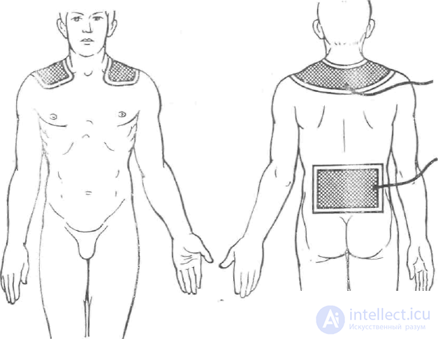
+Galvanization of the collar zone, see fig. The patient's position is lying down. One electrode in the shape of a shawl collar is placed on the upper part of the back so that its ends cover the shoulder girdle and clavicles up to the second intercostal space in front. The second electrode, with an area of 300 cm2, is placed in the lumbosacral region. The collar electrode is most often connected to the positive terminal of the galvanization device. With each procedure, the duration of exposure is increased by 2 minutes and the current strength by 2 mA, starting from 6 minutes and 6 mA, and increasing them up to 16 minutes and 16 mA. The course of treatment is 15-20 procedures.
Medicinal electrophoresis is a combined, i.e., simultaneous, therapeutic effect on the patient's body of a constant electric current and a medicinal substance, which enters the body with the current through intact skin or mucous membranes.
The possibility of introducing medicinal substances into the body by electric current through intact skin was first demonstrated in the work of the French researcher S. LeDuc (1908). The scientific basis for such research was S. Arrhenius's discovery of electrolytic dissociation, as a result of which not only neutral molecules but also oppositely charged ions are constantly present in electrolyte solutions. The proportion of molecules constantly dissociating into ions, as well as the number of molecules simultaneously formed from ions, depends on the nature of the substance, the concentration of the solution, its temperature, and, given identical conditions, on the nature of the solvent. The greater the dielectric permittivity of the solvent, the greater the degree of dissociation it causes and the more ions are present in the solution. For this reason, water, which has the highest dielectric permittivity among solvents, is most often used as the solvent. For substances that do not dissolve in water, aqueous solutions of dimethyl sulfoxide (DMSO), glycerin, ethyl alcohol, and others can be used as solvents that ensure dissociation of the molecules of the substance being dissolved. Their dielectric permittivity is, respectively, 48.9; 43; 25.8, etc., and that of water — 81 [Gerasimov Ya. I., Drevnich V. II, 1966].
Applying a constant electric current to a solution, as already noted, causes an oppositely directed movement of ions within it. Positively charged ions, which move toward the negative electrode (the cathode), are called cations, while negatively charged ions, moving in the opposite direction, are called anions.
The same movement of ions under the influence of direct current occurs in body tissues, which contain a considerable amount of dissolved salts. If a solution of some medicinal substance — an electrolyte — is placed between the electrodes and the body surface, then the ions of the medicinal substance it contains, being repelled from the like-charged electrode and moving toward the electrode of the other polarity, will penetrate into the body's tissues, which is what is used in medicinal electrophoresis.
The amount of medicinal substance entering the body with the current, and the pattern of its distribution in the tissues, which are of substantial importance in the mechanism of the therapeutic effect, are determined by the following factors and phenomena. Each ion in an electrolyte solution is surrounded by solvent molecules and by ions of opposite charge, forming what is called an atmosphere.
Ions moving under the influence of the current experience a retarding effect from solvent molecules, the ionic atmosphere, and ions moving in the opposite direction. The magnitude of the retarding effect of these factors depends on the concentration of the solution, its temperature, the viscosity of the medium, and other conditions. In aqueous solutions, at an electric field strength of 1 V/cm, the speed of ion movement is several centimeters per hour, for example for potassium ions (K+) — cm/h. Hydrogen ions (H+) have a much higher speed — 10 cm/h — and hydroxyl ions (OH-) — cm/h [Gerasimov Ya. I., Drevich V. P., 1966].
According to Faraday's first law, the amount of substance moving in a solution under the influence of current, or liberated at an electrode, is directly proportional to the amount of current passing through the solution.
According to Faraday's second law, moving the chemical equivalent of an ion in solution (the quotient of molar mass divided by valence), or liberating it at the electrodes, requires the same quantity of electricity — 9.65*104 C — known as Faraday's number, or Faraday's constant. The amount of substance liberated at the electrode or transported in the solution is determined by the formula:
m= M*I*t/n*F
where
m— the amount of substance liberated at the electrode or transported in the solution when a current I passes for a time t, F — Faraday's number, M — molar mass, n — the valence of the ion. However, in the tissues of the body, and to the greatest extent, the aforementioned patterns of electrokinetic processes undergo substantial changes. The skin is not only a significant mechanical barrier but is also an actively functioning organ with many functions aimed at maintaining homeostasis. In this process, fatty substances and salts excreted with sweat, and desquamating epidermis, are released. The desquamating horny plates of the epidermis and fatty substances further increase the already quite high resistance of the skin, and consequently reduce the total number of ions entering the body during electrophoresis. Elimination of the factors mentioned above is achieved by wiping the areas of skin on which the electrodes are to be applied with an alcohol-ether mixture, or by washing them with warm water and soap. The significance of other factors affecting the electrogenic entry of medicinal-substance ions into tissues has been studied in special research by V.S. Ulashchik (1976, 1979) and is discussed further below.
A certain influence on the penetration of medicinal substance ions into the body is exerted by the active acidity of the skin (pH). A shift of the skin reaction toward the acid side reduces its permeability for cations and increases it for anions. Alkalization of the skin leads to the reverse changes in its permeability.
The skin is more permeable to ions carrying a positive charge. Therefore, electrophoresis of substances with amphoteric properties (proteins and amino acids) is more advisable to perform from the anode, acidifying the working solution. An inverse relationship is observed between the amount of substance entering the tissues and the size of the ions and their valence. Greater penetration of ions through the skin is promoted by a reduction in ion hydration achieved by heating the working solutions.
Depending on the severity of the above-mentioned factors that hinder the penetration of ions into the tissues, the discrepancy between the actual transfer of substances during electrophoresis through the skin and the transfer of substances occurring in the solution, according to data from V.S. Ulashchik (1967), may range from 37.9 (lithium) to 85% (ganglerone).
The amount of medicinal substance ions entering the tissues with the current depends to a considerable degree on the choice of solvent. The best of these, providing the greatest dissociation and consequently the greatest amount of medicinal substance ions, is, as already noted, distilled water. Ionization of the water molecules themselves is negligible and can be disregarded. Physiological saline (0.85% sodium chloride solution) or a sodium bicarbonate solution should not be used as a solvent, since they contain a significant amount of ions which, possessing greater electrophoretic mobility, significantly limit the entry of the medicinal substance into the tissues. It goes without saying that the medicinal substance used for electrophoresis must not contain any impurities.
As for the concentration of the working solutions used for medicinal electrophoresis, some authors argue that the use of higher concentrations is advisable, while others do not.
The advisability of using higher concentrations of solutions in order to increase the amount of medicinal substance entering the tissues during electrophoresis. In studies by other authors (Kolker I.I., 1951, Abrekosov I.A., 1953), no significant increase was observed in the medicinal substances entering the body during electrophoresis of concentrated solutions. Studies conducted by Ulashchik to clarify this question showed that a noticeable increase in the content of the introduced substance in the skin, blood, and tissues, as well as in its pharmacological activity, occurs when the concentration of the working solution is increased to 2-3%. As the concentration is increased further from 3 to 5%, the entry of the medicinal substance into the body decreases, and at a concentration above 5% it stops entirely. S.C. Jacobsen and R.L. Stephen (1980) arrived at approximately the same results; studying concentrations of 1, 2, 4, and 10%, they found the 4% solution to be the most effective.
When a direct current passes through a section of the body on whose surface pads soaked in a solution of the medicinal substance are placed, through the skin, as through any porous membrane, not only electrophoresis but also diffusion and electroosmosis take place; according to the studies of I. Ipser (1958) as well as V.S. Ulashchik (1968), electroosmosis has no significant role in the transfer of medicinal substances into the body's tissues. According to Ulashchik's observations and calculations, electrophoresis accounts for 90-92% of the medicinal substance entering the body, diffusion for 5-8%, and electroosmosis for 1-3%. The same author established that an increase in current density from 0.03 to 0.1 mA/cm2 at the same duration, as well as an increase in procedure duration from 10 to 30 minutes, has no effect on the state of the skin's ionophoretic permeability with age.
Determination of the amount of substance entering the body during electrophoresis was carried out by I. Ipser. In the table he developed, data are presented for a number of substances; Ulashchik developed a more universal method for determining the amount of substance entering the body with the current, using a formula that takes into account the patient's age, electrode localization, the quantity of electricity, and the concentration of the solution. Calculations performed by the method mentioned show that during a one-minute procedure with a 1% solution at a current strength of 10 mA, 2-5% of the medicinal substance used in the procedure enters the body, for example 6.12 mg of ascorbic acid, 5 mg of novocaine. To speed up and simplify the calculations, tables have been compiled.
As for the depth of penetration of medicinal substances into the tissues during electrophoresis, most researchers note their penetration into the upper layers of the skin and the formation there of a so-called skin depot, from which the drugs, retained there for up to 3-20 days, diffuse into the blood and lymph and are distributed throughout the body. Thus V.S. Ulashchik (1976) found that after an electrophoresis procedure, radioactive substances were detected mainly in the epidermis and in the dermis proper. With repeated procedures, the concentration of the introduced substance increased in the deeper tissues.
P.D. Gadsby (1979), using an electron-microscopic research method, likewise established that electrophoretically introduced iron ions were detected only in the epidermis and the dermis proper. According to data from N.A. Barsukov, however, sulfonamides and antibiotics were detected not only in the subcutaneous tissue but also in the muscles at a depth of 2-3 cm, which is probably a result of hematogenous penetration.
In connection with the search for ways to increase treatment effectiveness, in recent times not only galvanic but also various pulsed currents have often been used for electrophoresis. Studies by G.K. Koltovich (1982, 1983) on the electrophoretic capacity of certain currents showed that the total amount of substances introduced into the body by diadynamic currents ranged from 20% (syncopal rhythm) to 86% (two-phase continuous current) in comparison with the amount of substance carried by galvanic current. Transfer of substance by fluctuating current was 7 times less. Rectified sinusoidal currents [SMT-I III. IV modes of operation (MO)] introduced about 60% of the substance into the tissues compared with galvanic current. In terms of electrophoretic activity, i.e., the transfer of substance in milligrams per coulomb (mg/C), the "syncopal rhythm" and "two-phase wave" of the diadynamic currents, as well as mode II of SMT, were not inferior to galvanic current.
The depth of substance penetration into the tissues, determined by the radiometric method, was found to be greater with mode II SMT, syncopal rhythm, and wave diadynamic currents than with galvanic current; with modes I, III, IV SMT and continuous diadynamic currents it corresponded to the depth obtained with galvanic current, while with fluctuating current and the "short" and "long periods" of diadynamic currents it fell short of the depth obtained with galvanic current.
Mechanisms of therapeutic action: Even upon initial consideration, it is evident that the therapeutic action of medicinal electrophoresis is made up of the influence of the direct current on the tissues and of the medicinal substance entering the body along with it. Orienting toward the action of the medicinal substance alone or predominantly during electrophoresis [Medvedev I.L., 1957; Goldman R.N., 1967] appears to us to be entirely unjustified. Galvanic current is an active irritant and stimulator of biological processes. Other direct currents used for electrophoresis possess no less pronounced a biological action. It is precisely for this reason that direct current cannot be regarded merely as a means of transport for delivering the medicinal substance into the body. Nor should medicinal electrophoresis be defined as a method of introducing drugs into the body by electric current. Such a definition not only fails to reflect the actual relationship among the processes taking place in medicinal electrophoresis, but also misleads those who are unfamiliar with the essence of the method.
Medicinal electrophoresis should not be spoken of, and still less written about, as a way of introducing medicinal substances into the body by electric current, not only because the direct current exerts a substantial influence on the tissues and the body, but also because, in comparison with other methods of administering drugs - intravenous, injection, inhalation, and others - only an insignificant amount of the medicinal substance enters the body despite the laboriousness of the procedure - 2-5% of that used in the procedure. It is hardly possible to count on a therapeutic effect from such a small amount of an individual medicinal substance taken alone. It would, however, be a mistake to disregard such small doses of medicinal substances in combination with the action of the current. Nor can the action of electrophoresis be regarded as a simple sum of the influence of the current and the medicinal substance. Electric current, by bringing the tissues, including the receptors, into a state of heightened activity and exciting them, makes them more sensitive to the action of drugs. This is also confirmed by the experiments of V.S. Ulashchik (1976). In this process the current may not only enhance but also weaken the action of drugs and alter their pharmacodynamics. There is likewise no doubt that medicinal substances can influence the action of the current, enhancing or weakening it. For the justified therapeutic application of electrophoresis of a medicinal substance not yet studied, it is not enough to rely solely on theoretical premises. Physicochemical studies are required, as well as experimental-clinical verification of the biological action and effectiveness of the technique in comparison with the action of the current alone.
The slow entry, accumulation in the skin depot, and subsequent diffusion into the lymph and blood flow of most substances of moderate potency, together with the active influence of the current, allow us to consider the action of the direct current predominant in the method of medicinal electrophoresis. This is confirmed by the facts of approximately equal effectiveness when the same technique is applied for galvanization and for electrophoresis of a number of substances, for example "transcardiac" electrophoresis of magnesium compared with galvanization. The exception is electrophoresis of potent substances. Thus, in experiments of 1908 the action of strychnine and cyanide compounds was predominant. In electrophoresis, the action of histamine, adrenaline, and other substances with a pronounced specific effect on the body comes to the fore.
In prescribing medicinal electrophoresis, along with taking into account the action of the current and the drug, one also proceeds from the features inherent to this method. These include:
- the gradual accumulation of the medicinal substance in the epidermis, as well as in the dermis proper, its retention there for several days (depot) with a course of treatment. This circumstance is advisable to use in those clinical situations where it is necessary to concentrate the action of the current and the medicinal substance on some limited area of tissue, for example on a joint, in the area of an injury, a scar, etc. The presence of certain medicinal substances in the skin depot can exert a reflex-segmental influence on the central nervous system and internal organs. A general reflex action is also possible - ionic reflexes according to Shcherbak.
- continuous entry of the medicinal substance into the bloodstream from the depot over a long period of time, which is advisable to use in chronic pathological conditions, when there is no need for large, so-called loading doses, for example electrophoresis of iodine in hyper- or dysthyroidism. The entry of medicinal substances into the blood can be enhanced by combining electrophoresis with inductothermy, or by combining preliminary (30-90 min beforehand) exposure to ultra-high-frequency electromagnetic oscillations [Klimovskaya I.G., 1979], ultrasound [Ulashchik V.S., 1979], or infrared and visible radiation.
- absence of the side effects that occur when preparations are administered by injection or per os;
- entry of the medicinal substance into the body in the form of ions, i.e., in an actively acting form.
As already noted, recently, with the aim of enhancing one or another component of the therapeutic action, rectified sinusoidal modulated or diadynamic currents have been used. In this case SMT enhances the local anesthetic action during electrophoresis of a novocaine-adrenaline mixture, while diadynamic currents prolong it. The use of nicotinic acid electrophoresis with modes I and IV SMT or continuous diadynamic currents produces a more significant and prolonged enhancement of regional blood flow compared with the use of galvanic current (Koltovich G.K., 1982, 1983).
Medicinal electrophoresis has been used in medical practice for more than 50 years. However, it has not become equally widespread everywhere. Thus, of 302 physiotherapy departments surveyed in the USA, electrophoresis was used in only 79 of them in 1971, and in only 6 of those in more than 5% of patients.
The main areas of its application were diseases of the skin, the organs of the ear, throat, nose, and oral cavity [Bonne O.C,. 1981]. The listed areas of application indicate that this method is used only for local effects.
In our country, electrophoresis accounts for 20-25% of all physiotherapy procedures [Ulashchik V.S., 1976]. Such widespread use of it can be explained by the simplicity and accessibility of the method, as well as by its reliance not only on local but also on reflex action.
The analysis presented above of the physical processes underlying medicinal electrophoresis shows that even with classical electrophoresis techniques, in which the electric current cannot enter the body without passing through the medicinal preparation, the amount of substance ions penetrating into the body is extremely small (2-5% of that placed on the pad). If, however, the electrodes are positioned on the surface of the body while the medicinal substance is administered into the body intravenously, intratracheally, into the uterus, rectum, or other organs and tissues, or is applied to the surface of the body but not under the electrodes, then no electrophoresis or other movement of this medicinal substance under the influence of the current can occur. Therefore, the terms "intratissue," "intraorgan," "intracerebral electrophoresis," or "electrodragging" should be regarded as not corresponding to the essence of the processes taking place during the procedures mentioned.
Indications for the therapeutic use of medicinal electrophoresis are determined by the previously listed features of the action of this method on the body. Given the leading role of the current in this method, the greatest density of which and the reactions it causes arise in the tissues under the electrodes, the main indications for medicinal electrophoresis should be local and regional processes. The medicinal substance is likewise selected based on these same considerations. A general action of the medicinal substance can be counted on mainly in functional vegetative-vascular disorders and conditions in which a microdose of the medicinal substance is sufficient, for example iodine in hyper- or dysthyroidism.
Procedure technique. For medicinal electrophoresis, the same electrodes are used as for galvanization. Most often this is a metal plate or graphitized fabric connected by a wire to the terminals of the apparatus, and a hydrophilic cloth pad. In order to make the ions of the medicinal substance move into the body, the substance is placed in the path of the current in such a way that the current cannot bypass it. To do this, 1-2 layers of filter paper, sized to match the cloth pad, are placed on the surface of the body. On the filter paper is placed a cloth pad moistened with water and well wrung out, and on top of that a metal plate. The electrode is then fixed in place. The filter paper under the second electrode is moistened with tap water or, if ions of the other polarity need to be introduced, with a solution of the medicinal substance.
When performing cavity procedures, the electrode inserted into the cavity, for example into the nose, is wrapped in a layer of cotton wool soaked in a solution of the medicinal substance. In some cases, the cavity is filled with a solution of the medicinal substance and the electrode is then inserted into it in such a way as to prevent contact of the metal part of the electrode with the body. For example, in electrophoresis of the prostate gland area, the rectal ampulla is filled with a solution of the medicinal substance and an electrode connected to the corresponding terminal of the apparatus is inserted. The second electrode (a plate electrode) is placed above the pubis. In electrophoresis of the ear, with the patient lying on their side, a solution of the medicinal substance is instilled into the auditory canal, filled with a cotton swab, and a cloth pad and the electrode's conductive plate are placed on top of the swab. The second electrode is placed in the cervico-occipital region or on the opposite shoulder.
Ions of medicinal substances are introduced from the electrode of like polarity. For example, metal ions and most alkaloids are introduced from the positive electrode, while ions of acid radicals and metalloids are introduced from the negative electrode.
The situation is more complicated with the electrophoresis of proteins, which are amphoteric polyelectrolytes. Their solutions for electrophoresis must have a certain pH value which, without disturbing the protein's activity, would promote its optimal migration, i.e., would differ, as far as possible, from the isoelectric point. For the electrophoresis of proteins one must use either already developed solvent recipes, or conduct preliminary studies to clarify the influence of direct current on the properties of the protein in various solvents. V.S. Ulashchik (1979), on the basis of studies conducted, proposes using acidified (introduced from the anode) or alkalized (introduced from the cathode) distilled water instead of buffer solutions. It should be kept in mind, however, that because of the large size of protein molecules, only certain proteins can be used for electrophoresis - hyaluronidase and its compounds, trypsin, fibrinolysin, and so on. In this case their penetration into the tissues is quite insignificant and shallow (only into the most superficial layers of the epidermis).
Amino acid molecules are smaller in size. They penetrate the skin with the current more readily. However, the choice of solvent must be made in the same way as for protein substances - must be made in the same way as for protein substances - with regard for the isoelectric point, bearing in mind that amino acids can be neutral, basic, or acidic. In acidified solutions amino acids migrate from the anode, and in alkalized solutions from the cathode. Among the amino acids used for electrophoresis are histidine, glutamic acid, methionine, and others.
Detailed information on the electrophoresis of individual substances is contained in the monographs of A.P. Parfenov (1973) and V.S. Ulashchik (1979).
In conclusion, it should be noted that when a direct current passes through a given part of the body, ions not only enter the body but are also removed from it. The process of ion removal, called elimination, has not found practical application because only a very small amount of substance is removed from the body. Moreover, it is not the ions that need to be removed from the body, for example ions of toxic substances, that are eliminated, but predominantly those located in the superficial tissues under the electrode that possess greater migratory mobility.
+Sometimes a medicinal substance is introduced into the body by one method, for example per os, intravenously, intramuscularly, or intratracheally, and at the same time the site of the drug's entry, for example the projection of the lungs, is exposed to direct current with the electrodes placed on the surface of the body, in the expectation that so-called intratissue electrophoresis will thereby take place. The physical processes discussed in this chapter, as already noted earlier, give no grounds for counting on migration of a medicinal substance introduced inside the body. With the combinations mentioned, an activation of blood circulation occurs in the tissues under the electrodes and, to a lesser extent, in the tissues of the metamere belonging to the same segment of the spinal cord as the areas under the electrodes.
Franklinization is a therapeutic method in which the patient's body, or individual areas of it, are exposed to a constant high-voltage electric field. Franklinization (electrostatic shower) is one of the oldest methods of electrotherapy. The electric field can reach 50 kV. With local exposure, 15-20 kV.
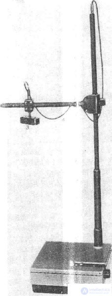
The franklinization procedure is carried out so that the patient's head (in general exposure) or another part of the body (in local exposure) serves, as it were, as one of the plates of a capacitor, while the second is an electrode suspended above the head, or positioned above the site of local exposure at a distance of 6-10 cm.
Since the resistance of the body is small compared with the resistance of the air, all the voltage generated by the apparatus drops across the air gap between the patient's body and the electrode. However, under such high voltage, ionization of the air occurs beneath the points of the needles fixed on the electrode, with the formation of air ions, ozone, and nitrogen oxides. In the tissues located opposite the electrode, under the influence of the voltage, polarization of molecules occurs with the appearance of microcurrent in areas with good electrical conductivity. There is likewise a change in the usual ratio of ions in the tissues. Inhalation of ozone and air ions causes reactions of the vascular system. After a brief spasm of the vessels, dilation of the capillaries occurs, not only in the superficial tissues but also in the deep ones. As a result of this, metabolic processes are stimulated. In addition, processes of regeneration and restoration of function are stimulated, the blood supply to the brain and its membranes is improved, which leads to a reduction in headaches and an increase in arterial pressure. An increase in vascular pressure, and a slowing of the pulse.
Procedure technique. Exposure to static electricity is carried out with the patient seated on a wooden chair or couch. In doing so, the chair or couch should not be placed near or adjacent to grounded objects (even shielded ones). Before the procedure, all metal objects must be removed from the patient's hair and clothing pockets, in order to avoid deformation of the electric field and undesirable amplification of the effect in unforeseen locations. During general exposure, the patient, in light clothing, sits on the chair with the feet placed on the foot electrode. Rubber footwear is removed for this. The second electrode, in the form of a "spider" with points directed downward, is suspended above the patient's head at a distance of 12-15 cm from its surface. The field strength is set at a level of 40-50 kV. The duration of the procedures, carried out daily or every other day, is 10-15 min, with a course of treatment of 10-15 procedures. During the procedures, patients must not touch any objects or touch anyone.
When performing local exposure, the wound or ulcer surface must be cleared of pus, rejected tissue masses, and crusts, treated with the appropriate medicinal solutions, and dried with a sterile wipe. The patient is placed in a comfortable position. Above the area of exposure, at a distance of 5-7 cm from the surface of the wound or ulcer, an electrode is fixed on a bracket screwed to the couch or chair. A foot electrode is placed under the corresponding part of the body. For local exposure, a voltage of 10-20 kV is used. The duration of the procedures, carried out as a rule during dressing changes (every 2-3 days), is 10-15 min, with a course of treatment of 10-15 exposures.
+General franklinization is indicated for functional disorders of the nervous system: neurasthenia with asthenic syndrome, migraine, insomnia, physical and mental fatigue and overexertion, and residual effects of arachnoiditis. Local franklinization is used for trophic ulcers, infected wounds with a sluggish course, burns, local itching, and paresthesias.
+Blood diseases, malignant tumors, pronounced atherosclerosis of the cerebral vessels, impaired cerebral blood supply, febrile conditions, pregnancy, active pulmonary tuberculosis.

Comments