
Lecture
1-1. GENERATION OF X-RAYS
As already noted, X-rays arise at the anode of an X-ray tube when electrons moving from the cathode to the anode under the action of the accelerating field are decelerated. During deceleration, the following processes take place:
a) elastic collisions with atoms and ionization in the outer electron shells; in these processes the kinetic energy of the electron is gradually converted into thermal energy;
b) ionization in electron shells close to the atomic nucleus, which leads to the appearance of the X-ray characteristic spectrum, consisting of separate spectral lines;
c) sharp deceleration of the electron in the field of the nucleus, as a result of which a continuous X-ray bremsstrahlung spectrum arises with a sharp boundary on the short-wavelength side.
Almost all of the kinetic energy of the decelerating electrons is converted into thermal energy. Only an insignificant fraction — of the order of tenths of a percent or a few percent (depending on the voltage) — is converted into the energy of X-ray radiation. Therefore, cooling of the anode must be provided in the tubes. Heating of the anode limits the permissible power of the tube.
X-ray radiation is characterized by intensity and hardness. Intensity is understood as the amount of radiation energy incident on a unit surface per unit time. Hardness is understood as the relative penetrating power
of the rays; the harder the rays, the greater their penetrating power. In the case of homogeneous (i.e., single-wavelength) radiation, hardness can be uniquely characterized by this wavelength; the shorter the wavelength (i.e., the more energetic the X-ray photons), the greater the penetrating power of the radiation. Assessment of the hardness of a continuous X-ray spectrum is more complex and will be discussed further below.
The specific properties of the characteristic spectrum are used only in certain methods of structural analysis and in spectral analysis. The characteristic spectrum has significantly less energy than the bremsstrahlung spectrum. Therefore, when using the entire radiation, it can be assumed as a first approximation that the effect of X-rays is due only to the bremsstrahlung spectrum.

a) Bremsstrahlung spectrum. All electrons at the surface of the anode have the same kinetic energy. The electron acquires this kinetic energy while moving in the accelerating field between the cathode and the anode. Therefore, it is equal to:
(1-1)
where e — is the charge of the electron, equal to 4,802 • 1010 esu, and Ua~ the anode voltage, i.e., the voltage between the electrodes of the tube (the initial kinetic energy that the electron possesses upon leaving the cathode can be neglected owing to its smallness). However, the photons of the X-ray bremsstrahlung spectrum will have different energies. This can be explained by the fact that: 1) an electron may undergo sharp deceleration after having lost part of its energy in elastic collisions; 2) during sharp deceleration an electron may partially retain its kinetic energy. As a result, the radiation generated by the tube will contain photons with all possible energies up to a certain maximum, characterizing the complete conversion of the electron's entire kinetic energy, stored in the accelerating field, into the energy of X-ray radiation.
The relationship between the photon energy A and the wavelength A is given by the formula

where h — Planck's constant, equal to 6,62- 10~2 erg-cm, and c — the speed of light, equal to 3 • 1010 cm/sec. Thus, the photon with maximum energy corresponds to the minimum wavelength. Equating formulas (1-1) and (1-2), we see that the minimum wavelength, like the maximum photon energy, is determined by the anode voltage. Substituting numerical values, we obtain the formula:
λmin =12,4/Ua
If the voltage in this formula is expressed in kilovolts, then the wavelength
is obtained in angstroms (A);
1 A = 10 8 cm. Formula (1-3) can also be used to convert from wavelength to photon energy, if the latter is measured in kiloelectronvolts (keV). This unit of energy has become widely used in application to elementary particles.
The distribution of energy in the bremsstrahlung spectrum is shown in Fig. 1-1. Here the abscissa is the wavelength of the radiation, and the ordinate is the intensity of radiation at a given wavelength (the so-called intensity density in the spectrum) Jx. The curves in Fig. 1-1 were taken at the same anode current and different anode voltages. Fig. 1-1 shows that as the voltage increases, there is a shift toward shorter wavelengths not only of the minimum wavelength, but also of the intensity maximum, i.e., of the entire spectrum as a whole. As a result, increasing the voltage causes an increase in the hardness (penetrating power) of the bremsstrahlung rays.
The area bounded by the curve Jλ = f(λ) and the abscissa axis represents the total intensity of the radiation:
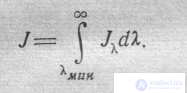
+Since this area increases as the anode voltage increases, it follows that the intensity of the radiation also increases as the voltage rises. The increase in radiation intensity with increasing voltage and a constant anode current (i.e., with a constant number of electrons decelerated at the anode of the tube per unit time) occurs owing to the increase in the kinetic energy of each individual electron. Conversely, an increase in the anode current at a constant anode voltage increases the number of electrons decelerated at the anode, while leaving the kinetic energy of an individual electron unchanged. Naturally, the hardness of the radiation does not change in this case, while the intensity increases in proportion to the number of electrons, i.e., in proportion to the anode current.
X-ray radiation is a type of electromagnetic oscillation arising from the sharp deceleration of accelerated
electrons at the moment of their collision with the atoms of the anode material of the X-ray tube, or during the rearrangement of electron
shells of atoms.
In their physical nature, X-rays do not differ in any way from other types of electromagnetic oscillations
(visible light rays, infrared, ultraviolet rays, radio waves, etc.). The difference lies only in the wavelength.
The place occupied by X-rays among other electromagnetic radiations is shown in Table 1.
X-ray radiation is generated in an X-ray tube, which is a device consisting of
a glass envelope and two metal electrodes, the cathode and the anode (anti-cathode). Inside the envelope,
a high vacuum is created. The cathode is connected to the negative, and the anode to the positive pole of the high-
voltage source.
Wavelengths of Various Types of Electromagnetic Radiation
Table 1
|
Type of radiation |
Wavelength |
|
Cosmic rays Gamma rays of radioactive elements X-rays Ultraviolet rays Visible light rays Infrared rays Radio waves
|
5*10-5 – 1*10-3 1*103 – 3*10-3 3*10-3 – 1,5 1.5 – 100 400 – 700 400 – 0,15 cm
0,15 cm – 30 km |
A voltage of about 10 V is applied to the cathode filament. When current flows in the filament circuit, the cathode begins to emit free
electrons (electron emission), which form an electron cloud around it (Fig. 1,a). Under the influence of the high
voltage between the anode and the cathode, the electrons rush toward the positively charged anode. The tube also has a
focusing device, which directs the electron stream to a single point – the focal spot of the anode. Upon collision
of the electrons with the anode, as a result of sharp deceleration, the kinetic energy of the electrons is converted into thermal
energy and the energy of X-ray radiation (Fig. 1,b).
Since the formation of X-rays in the variant considered is associated with the deceleration of the electron stream,
this type of radiation is called bremsstrahlung.
The intensity of X-ray radiation is proportional to the current strength, the square of the voltage on the tube, and the atomic number
of the anode substance. It can be calculated using the formula
F=K*Z*V2*I,
Where F is the intensity of X-ray radiation, V is the voltage, I is the current strength in the X-ray tube, Z is the atomic number
of the anode substance element, K is the coefficient of proportionality (K=10-9*V).
The magnitude of the current through the tube (in mA) depends on the number of free electrons, the source of which is the filament
of the cathode. By changing the voltage in the filament circuit of the tube, the intensity of X-ray radiation can easily be regulated.
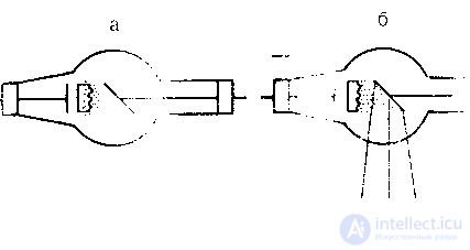
a - electron emission, b - generation of X-ray radiation.
Thus, if the current through the tube is increased from 2 to 4 mV, the intensity of X-ray radiation will increase by a factor of 2. If, however,
the voltage on the tube is doubled, the radiation intensity will increase by a factor of 4. However, in this case, not only
the quantity but also the quality of the X-rays will change, which is characterized by the energy of the radiation.
Energy is expressed in joules, but in electrical engineering another unit is also adopted – the electronvolt (eV). The electronvolt is energy.
That an electron acquires in an electric field with a potential difference of 1 V. This is a very small quantity. It
corresponds to an energy of 1,6*10-19 joules. Therefore, multiple units are used – the kiloelectronvolt (keV= 103 eV)
As the high voltage (the potential difference across the electrodes of the tube) increases, the energy of the radiation increases and
the wavelength of the X-rays decreases. Short-wave radiation is commonly called «hard». It has
greater penetrating power than long-wave radiation – «soft».
In addition to bremsstrahlung X-ray radiation, there is also so-called characteristic radiation. It is formed
owing to the fact that accelerated electrons penetrate deep into atoms and knock electrons out of the inner shells.
The vacated places are occupied by electrons from higher levels. In this process, the atom emits photons of characteristic
X-ray radiation, so named because it characterizes the substance of the anode of the X-ray tube.
Unlike bremsstrahlung radiation, which has a continuous spectrum, characteristic X-rays have a spectrum
that is line-type (discrete), typical of the atoms of each chemical substance.
1) The use of X-ray radiation in medicine for diagnosis and treatment is based on its ability to:
penetrate various substances, including organs and tissues of the human body that do not transmit rays of visible
light;
cause fluorescent glow in certain chemical compounds (activated zinc and cadmium sulfides, crystals
of calcium tungstate). This property is the basis for X-ray fluoroscopy, as well as the use of intensifying screens
in radiography.
2) Exert a photochemical effect (decompose compounds of silver with halogens) and cause changes in photographic
properties (including of radiographic film). This property is the basis for obtaining X-ray images;
4) It causes physiological and pathological changes (depending on the dose) in irradiated organs and tissues
(exert a biological effect). This property is the basis for the use of X-ray radiation in the treatment of
tumor and certain non-tumor diseases. However, with insufficiently controlled irradiation at large doses
the development of acute or chronic radiation sickness or radiation injuries is possible;
5) Transfer the energy of the radiation to atoms and molecules of the surrounding medium, causing their excitation, as well as decay into
positive and negative ions - the ionizing effect. Under certain conditions, between the ionization
effect and the duration of irradiation there is a direct relationship. This makes it possible, by assessing with the aid of special
instruments (dosimeters) the degree of ionization of the air, to determine the quantity and quality of X-rays,
used for diagnosis and therapy.
When X-rays interact with matter, absorption of photon energy occurs (a photon is a particle
of electromagnetic radiation that causes ionization and excitation of atoms and molecules). The mechanism of interaction
of X-rays with matter depends mainly on their energy. Three main types of energy transfer
+of electromagnetic radiation to matter are distinguished: the photoelectric effect, the Compton effect, and pair production (Fig. 2):
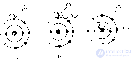
a — photoelectric effect; b— Compton effect; c — pair production.
In the photoelectric effect, the energy of the incident quantum is fully expended on knocking out an orbital electron
of the atom it encounters and imparting kinetic energy to it. As a result, the atom that has lost an electron becomes a
positive ion. The ejected electron (photoelectron), in the process of its motion, in turn causes ionization of
other atoms. If the photon energy is insufficient for ionization, interaction of the photoelectric type may be accompanied by
excitation of atoms without electron ejection. In other cases, the transition of electrons from outer shells to inner shells
of the atom ends with the emission of a quantum of characteristic radiation. The photoelectric effect is observed at relatively low
radiation energies (10-50 keV);
Compton scattering (the Compton effect) occurs at higher levels of radiation energy (100—200 eV
and above). This type of interaction of radiation with matter differs from the photoelectric effect in that the ejected electron
is not given the entire energy of the incident quantum, but only part of it. The ejected electron, which received the name Compton electron, and
the recoil electron, possessing sufficient energy, take part in secondary ionization processes. On the other hand,
the incident quantum, having lost part of its energy in ejecting the electron and imparting motion to it,
may nevertheless retain a reserve of energy sufficient for new acts of ionization.
Pair production is observed during the interaction of high-energy radiation (above 1,02 MeV) with the nuclei
of heavy atoms. In this process, a pair is formed: an electron and a proton.
X-RAY IMAGE
FORMATION AND BASIC PROPERTIES OF THE X-RAY IMAGE
The X-ray image is formed on radiographic film, the screen of an X-ray machine, the output screen of an image intensifier tube, or on the screen of an X-ray television device, and, in essence, represents a complex combination of numerous shadows differing from one another in size, shape, structure, and optical density. Analyzing this picture, the radiologist must draw a conclusion about the normal condition of the organs being examined or about the presence in them of one or another pathological change. To correctly solve this problem, it is necessary that the radiologist and the X-ray technician be familiar with the features of the X-ray image and be able to correctly assess the role of the various factors that influence its informativeness.
So how is an X-ray image formed?
First it should be emphasized that the X-ray image differs substantially from the photographic image, as well as from the ordinary optical image created by visible light. Thus, electromagnetic waves of visible light, emitted by some bodies or reflected from them, upon entering the eye, produce visual sensations that create an image of the external appearance of the object. Likewise, a photographic picture reflects only the external appearance of the object being photographed, usually on a reduced scale. The X-ray image, on the contrary, reproduces the internal structure of the body under examination. Its formation is associated with the uneven absorption of radiation by different tissues.
As is known, the absorption of X-rays, besides their energy, is determined by the atomic composition, density, and thickness of the object.. The heavier the chemical elements making up the tissues, and the greater their density and layer thickness, the more intensely the X-ray radiation is absorbed. And, conversely, tissues consisting of elements with a low atomic number usually have a low density and absorb to a lesser degree ; X-rays. Table 2 gives data on the density of various tissues and media making up the human body.
Table 2
Density (g/cm3) of various tissues and media making up the human body.
|
Tissues and substances |
Density |
|
Air Fat Water Muscles Cartilage Bones |
|
As can be seen from this table, the human body consists of substances having different densities.
It has been established that if the relative absorption coefficient of medium-hardness X-ray radiation by water is taken as 1,0 then for air it will be 0,01; for fatty tissue – 0,5; calcium carbonate – 15,0; calcium phosphate – 22,0.
In other words, X-rays are absorbed to the greatest degree by bones, to a considerably lesser degree—by soft tissues (especially fatty tissue), and least of all by tissues containing air.
The uneven absorption of X-ray radiation in the tissues of the anatomical region under examination causes the formation, in the space beyond the object, of a modified or non-uniform beam of X-rays (the exit dose or the dose beyond the object). In essence, this beam contains within it an image invisible to the eye (the image in the beam). Acting on a fluorescent screen or radiographic film, it creates the familiar X-ray image.
Thus, it is evident that the formation of the X-ray image is based on the unequal absorption of X-rays in the organs and tissues under examination. This is the so-called absorption law of X-ray differentiation. Its essence lies in the fact that any object (any anatomical structure) on a radiograph or fluoroscopic screen can give rise to a separate shadow only if it differs from the objects (anatomical structures) surrounding it in atomic composition, density, and thickness.
Depending on the ratio of dark and light areas, the X-ray image can be positive or negative. A positive image is formed in fluoroscopy. On the fluoroscopic screen, the lightest (brightest) areas are those corresponding to anatomical structures that are «transparent» (having greater density and thickness) to X-rays. Such structures primarily include air-containing lung tissue, the paranasal sinuses, the intestine, containing gas, as well as soft tissues, especially fatty tissue. Conversely, anatomical structures that intensively absorb X-rays (bones, various kinds of calcifications, massive formations, etc.) create dark areas on the screen. Thus, when the chest is examined by fluoroscopy against the background of transparent (light) air-containing lung tissue, dark areas caused by the tissues of the ribs, the roots of the lungs, the heart, and the large vessels are clearly identified.
On radiographs, the ratio of dark and light areas is reversed: light areas correspond to anatomical structures that absorb radiation the most, while dark areas correspond to the more transparent areas of the object under examination. Such an image is negative. To avoid confusion in describing radiographs, the relationships characteristic of fluoroscopy are used as the basis. Therefore, light areas on negative radiographs are conventionally called «dark areas», and dark areas «light areas».
The X-ray image is created by the beam of X-rays that has passed through the object under examination. Along its path, each ray naturally crosses a multitude of points, each of which, to one degree or another (depending on the atomic composition, density, and thickness), absorbs its energy. In this case, the total attenuation does not depend on the spatial arrangement of the individual points absorbing the radiation. This regularity is seen from Fig. 3. Despite the different arrangement of tissues within the volume under examination, all points that together caused the same attenuation of the X-ray beam, when examined in a single projection, are displayed on the plane in the form of shadows of the same intensity.
This example demonstrates that the X-ray image is planar and summational.
The planar and summational nature of the X-ray image can give rise not only to summation, but also to a kind of subtraction of shadows. Indeed, if there are areas of compaction and areas of rarefaction along the path of the X-rays, then the increased absorption in the first case is compensated by the decreased absorption in the second (Fig. 4).
Therefore, in an examination in a single projection, it is not always possible to distinguish a true compaction or rarefaction in a given organ from the summation or, conversely, the subtraction of shadows located along the path of the X-ray beam.
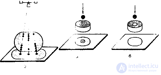
3. Diagram of the formation on the radiograph of an identical summed image of several points with different spatial arrangement in the object under examination (after V. I. Feoktistov)
4. Schematic representation of the effect of summation (a) and subtraction (b) when imaging in a single projection.
5 6
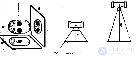
5. Diagram of the formation of summed (a) and separate (b) images of two shadows when imaging in two mutually perpendicular projections.
6. Schematic representation of the relationship between the tube focus–object distance and the projective magnification of the X-ray image of one and the same object. As the tube focus–object distance (F and F1) increases, the projective magnification of the X-ray image (AB and A1 B1) decreases.
From this follows a very important rule of X-ray examination: in order to obtain a differentiated image of all the anatomical structures of the region under examination, one should strive to take images in at least two (preferably three) mutually perpendicular projections – frontal, lateral, and axial – or to rotate the patient during fluoroscopy (Fig. 5).
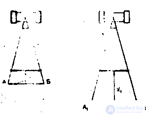
7. Schematic representation of the relationship between the object—image receiver distance and the projective magnification of the X-ray image of one and the same object. As the distance object—image receiver (X and X1) increases, the projective magnification of the X-ray image (AB and A1 B1) increases.
As already mentioned, X-ray radiation propagates from its place of origin (the focal spot of the anode) in the form of a diverging beam (cone), which leads to magnification of the X-ray image. The degree of projective magnification depends on the spatial relationships between the X-ray tube, the object under examination, and the image receiver (radiographic film, screen, selenium plate, etc.). This relationship is expressed as follows: the smaller the distance from the tube focus to the volume under examination, and the greater the distance from the object to the image receiver, the more pronounced the magnification of the X-ray image. Conversely, as the focal distance increases, the dimensions of the X-ray image approach the true dimensions (Figs. 6,7).
Consequently, in cases where it is necessary for the dimensions of the X-ray image to be close to the true dimensions, the object under study should be brought as close as possible to the cassette or fluorescent screen, and the tube should be moved as far away as possible. In fulfilling this latter condition, the power of the X-ray unit must be taken into account, since the intensity of the radiation varies inversely with the square of the distance. In practical work the focal distance is usually increased to a maximum of 2-2.5 cm (teleroentgenography). Under these conditions the projection magnification of the X-ray image is minimal. For example, the increase in the transverse size of the heart when imaging in the direct anterior projection amounts to only 1.0-1.5 mm.
Although an X-ray image is in principle always magnified, under certain conditions a projection reduction of the object under study is observed. Such a reduction usually concerns the image of planar formations or of structures having an elongated,
a b
8. Diagram of the features of the X-ray image of a section of a blood vessel (a) and a bronchus (b) depending on the position of their main axis relative to the central beam of X-ray radiation and to the image receiver (according to L. D. Lindenbraten).
elongated shape (bronchi, vessels), if their main axis is not parallel to the plane of the image receiver and not perpendicular to the central beam of X-ray radiation. The shadows of bronchi, as well as of vessels or of any other objects of elongated shape, have maximal dimensions in cases where their main axis is parallel to the cassette and perpendicular to the direction of the central ray. As the angle formed by the central ray and the long axis of the object under study approaches or increases, the dimensions of the shadow of the latter gradually decrease. In the orthograde projection (along the course of the central ray) a blood-filled vessel, like any linear formation, is displayed as a punctate homogeneous shadow, while the bronchus has the appearance of a ring (Fig. 8). A combination of such shadows is usually determined on radiographs or on the screen of the X-ray unit during fluoroscopy of the lungs. Unlike the shadows of other anatomical structures (thickened lymph nodes, dense focal shadows), when turned they acquire a linear character.
The formation of the X-ray image of planar formations occurs in an analogous way (in particular, in interlobar pleurisy). The shadow of a planar formation has maximal dimensions in cases where the central ray is directed perpendicular to the plane under study and to the film. If, on the other hand, the central ray slides along the planar formation (orthograde projection), then it is displayed on the radiograph or screen as an intense linear shadow. In all the examples considered, the central beam of X-ray radiation passes through the center of the object under study and is directed at the center of the film (screen) at an angle to its surface. This is what is usually sought in X-ray diagnostics. However, in practical work the object under study is often located at some distance from the central ray, or the cassette with the film or the screen is positioned not at a right angle to it (oblique projection). In such cases, deformation of its image occurs owing to the uneven magnification of individual segments of the object.
Thus, bodies having a spherical shape become elongated predominantly in one direction and take on the shape of an oval. (Fig. 9). Such distortions are most often encountered when examining certain joints (the heads of the femur and humerus), as well as when performing intraoral dental radiographs
In order to overcome projection distortions, in each specific case an attempt is made to achieve optimal spatial relationships between the object under study, the image receiver, and the central ray. For this the object is positioned parallel to the film (screen), and the central ray is directed through its central section perpendicular to the film. If, for one reason or another (a forced position of the patient, a peculiarity of the structure of the anatomical region), it is not possible to give the object the necessary position, then normal imaging conditions are achieved by correspondingly changing the position of the tube focus and the cassette (Fig. 10).
The perspective of the X-ray image (the image of three-dimensional objects on a plane) differs substantially from the image of objects that is familiar to our eye. As is known, in ordinary visual perception the relief and the image of an object is achieved mainly because distant objects have smaller dimensions than those closer to us, and are partially or fully obscured by them. In addition, perception of volume is aided by the presence, under lateral illumination, of light and shadow (darker and lighter areas).
On a radiograph or on the fluoroscopic screen, details of the image located farther from the image receiver
always have larger dimensions and less distinct contours than structures adjacent to it. Thus, on a direct anterior chest radiograph the image of the posterior sections of the ribs has larger dimensions and less distinct contours than that of the anterior sections.
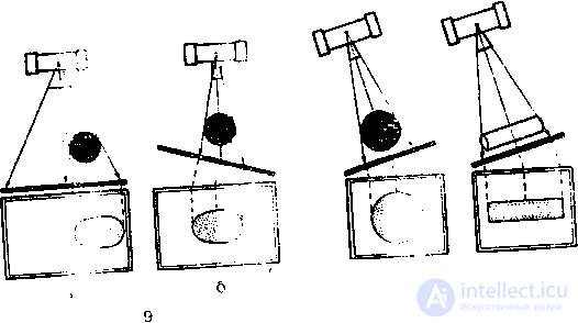
9. Diagram of various types of distortion of the X-ray image of a sphere when imaging with an oblique beam of X-ray radiation (a) and when the image receiver is positioned obliquely relative to the central beam of X-ray radiation (b),
10. Diagram of obtaining a correct image of objects of spherical (a) and elongated (b) shape when examining in an oblique projection. The image receiver is positioned in such a way that the central beam of X-ray radiation passes through the center of the object, perpendicular to the image receiver. In this case the longitudinal axis of the elongated object is positioned parallel to the plane of the image receiver
In addition, on a radiograph the posterior sections of the object under study are not obscured by the anterior sections, and the intensity of their display depends on the degree of absorption of X-ray radiation. Finally, owing to the above, the gradation of shadow intensity on radiographs has nothing in common with the distribution of light and shadow in visual perception of objects. Therefore, in the process of studying an X-ray image, a notion of volume can be obtained only through the simultaneous analysis of several images taken in different projections.
The intensity of the shadow of one or another anatomical structure depends on its radiolucency, i.e. its ability to absorb X-ray radiation. This ability, as has already been said, is determined by the atomic composition, density, and thickness of the object under study. The heavier the chemical elements comprising anatomical structures, the more they absorb X-ray radiation. A similar relationship exists between the density of objects and their radiolucency. The greater the density of the object under study, the more intense its shadow. This is precisely why, during fluoroscopic examination, metallic foreign bodies are usually easily identified, while it is very difficult to locate foreign bodies of low density (wood, various types of plastic, aluminum, etc.).
In most cases anatomical structures composed of heavy chemical elements have a high density. Thus, bones, which contain the heavy elements calcium and phosphorus, possess the greatest density among the tissues of the body and absorb X-ray radiation intensely; however, there are exceptions to this. In particular, it is known that air has considerably greater radiolucency than water. At the same time, nitrogen and oxygen (the chemical elements comprising air) are heavier than hydrogen and oxygen (the chemical elements of water).
Depending on density, it is customary to distinguish four degrees of transparency of media: air, soft-tissue, bone, and metal. Thus it is evident that in analyzing an X-ray image, which represents a combination of shadows of varying intensity, it is necessary to take into account the chemical composition and density of the anatomical structures under study.
However, it should be kept in mind that most tissues of the human body differ only slightly from one another in composition and density. Thus, muscles, parenchymal organs, brain, blood, lymph, nerves, various soft-tissue pathological formations (tumors, inflammatory granules), as well as pathological fluids (exudate, transudate) possess almost identical radiolucency. Therefore, the decisive influence on the intensity of the shadow of a given anatomical structure is exerted by a change in its thickness.
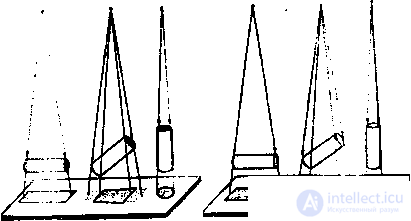
+It is known, in particular, that as the thickness of the body increases in arithmetic progression, the beam of X-rays behind the object (the exit dose) decreases in geometric progression, and even insignificant fluctuations in the thickness of the structures under study can substantially change the intensity of their shadows. As can be seen from Fig. 11, when imaging an object having the shape of a triangular prism, the sections of the shadow with the greatest intensity correspond to the maximal thickness of the object. Thus, if the central ray is directed perpendicular to one of the walls of the base of the prism, the shadow intensity will be maximal in the central section. As one moves toward the periphery, its intensity gradually

11. Schematic representation of the shadow intensity of various objects depending on shape, method of acquisition, and structure:
a, b – triangular prism, c – solid cylinder, d – hollow cylinder.
decreases, which fully reflects the change in thickness of the tissues located along the path of the beam of X-ray radiation (Fig. 11a). If, however, the prism is turned (Fig. 11, b) in such a way that the central ray is directed tangentially to one of the sides of the prism, then the edge section of the shadow corresponding to the maximal (in this projection) thickness of the object will have the maximal intensity. In a similar way the intensity of shadows having a linear or elongated shape increases in cases where the direction of their main axis coincides with the direction of the central ray (orthograde projection).
When examining homogeneous objects having a spherical or cylindrical shape (heart, large vessels, tumor), the thickness of tissues along the course of the beam of X-ray radiation changes very insignificantly. Therefore the shadow of the object under study is almost homogeneous (Fig. 11, c).
If, however, the spherical or cylindrical formation is hollow, then the beam of X-ray radiation passes through a greater volume of tissue in its peripheral sections, which causes the appearance of more intense areas of darkening, the so-called «edge rims» (Fig. 11, d). The latter are observed on radiographs of tubular bones, vessels with calcified walls, cavities with dense walls, etc.
It should be kept in mind that in practical work, for the differentiated perception of each individual shadow, the decisive factor is not the absolute intensity but the contrast, i.e. the difference in intensity between a given shadow and the shadows surrounding it. In this regard the physico-technical factors that influence the contrast of the image acquire important significance: radiation energy, exposure, the presence of a scatter grid, grid efficiency, the presence of intensifying screens, etc. Incorrectly chosen technical conditions of examination (excessive tube voltage, too high or insufficient exposure, low grid efficiency), as well as errors in the photochemical processing of films, reduce the contrast of the image and thereby have a negative effect on the differentiated identification of individual shadows and the objective assessment of their intensity.
+The application of ionizing radiation (IR) in the radiation therapy (RT) of malignant tumors is based on a profound understanding of the biological action of IR on various organs, tissues, and tumors, which represents an extremely complex process accompanied by certain morphological and functional changes in the irradiated tissue. In this process a combination of regressive phenomena with restorative ones is clearly traced, these being closely dependent on the absorbed energy and on the time elapsed after irradiation. Clear conceptions of these processes have served as the basis for the successful application of radiation for therapeutic purposes, as a means allowing the destruction of tumor tissue and the suppression of its growth, while at the same time avoiding irreversible post-radiation changes in the normal organs and tissues surrounding the tumor.
In the biological action of IR the first link is the absorption of radiation energy with subsequent interaction of it with the substance of the tissue, which proceeds over a very short time - fractions of a second. As a result of this interaction, in the cells of tissues and organs a whole chain of biophysical, biochemical, functional, and morphological changes develops, which, depending on specific conditions, unfold over various periods of time - minutes, days, years. During the interaction of radiation with matter, ionization and excitation of the atoms and molecules of the irradiated substance occur, and heat is generated. During irradiation, the processes of ionization and excitation arise only along the path of the ionizing particle.
As a result of the ionization of an atom or molecule, two ions arise, with positive and negative charge. Both ions are unstable, chemically active, and have a pronounced tendency to combine with central molecules, upon excitation of which the electronic configuration of the molecule changes, which can lead to the rupture of its molecular bonds. The breakdown products of the reacted molecules also turn out to be chemically active and, in turn, enter into chemical reactions with neutral molecules. The ionization of water molecules, which make up more than 80% of the organism, leads to its breakdown and to the formation of H+, OH, H2O2, H2; which possess considerable chemical activity and cause the oxidation of substances soluble in water.
Thus, the primary physical processes - ionization and excitation of atoms and molecules - lead to a chemical rearrangement of the irradiated molecules. In the primary mechanism of biological action a distinction is made between direct action (changes arising in the molecules of cells as a result of ionization or excitation) and indirect action (which combines all the chemical reactions proceeding with chemically active, but not ionized, products of dissociation of ionized molecules).
The processes of ionization and excitation are the triggering mechanisms that determine all subsequent changes in the irradiated tissues. The possibility of ionization depends on the size of the molecule: the larger its size, the greater the probability of its interaction with an ionizing particle. All the most important molecules have a large volume. An example is provided by DNA molecules, which take part in the transmission of heredity, in the processes of reproduction, and in the regulation of metabolism in the cell. Irradiation leads to the rupture of molecules and disruption of the structure of DNA. In an irradiated cell the processes of regulation and the activity of its individual components (membranes, mitochondria, etc.) are disrupted. The death of cells, even upon irradiation with large doses, can be spread out over a considerable time. Two types of cell death due to irradiation are distinguished: mitotic death (inactivation of the cell following irradiation after the first or a subsequent mitosis) and interphase death (death before the cell enters the mitotic phase).
The indirect action of radiation causes less severe disruptions, often reversible, but they encompass a larger number of molecules in a volume of tissue significantly exceeding the dimensions of the irradiation fields. An example of indirect action is the general reaction of the organism, leukopenia, which develops even in cases where the bone marrow is excluded from the irradiation zone.
The intensity of the reactions associated with the direct and indirect mechanisms of IR action depends, in addition to the initial state of the organism, on a number of physical and chemical factors. The physical factors include the dose and its rate - as these increase, the biological effect is intensified. The biological effect also depends on the quality of the radiation, which is characterized by LET and LPI, since the effect of irradiation is determined not only by the amount of absorbed energy but also by its macro- and microdistribution in the tissues.
Among the chemical factors influencing the biological effect, the influence of oxygen is the most pronounced. In the presence of oxygen a large quantity of chemically active radicals and peroxides arises, intensifying the oxidation processes in the irradiated tissues. The lifetime of primary radicals does not exceed fractions of a second, while the newly formed oxidants exist for a long time. In this process chain reactions can arise, and the chains that arise are longer the higher the oxygen content. Oxygen can enter into reaction with certain ionized molecules and promote their alteration, which might not manifest itself in the absence of oxygen. By increasing the intensity of the primary reactions developing under the influence of irradiation, oxygen raises the radiosensitivity of the cell, and this increase occurs instantaneously following an increase in oxygen content. The oxygen effect is most pronounced for radiation of an electromagnetic nature; it is higher for fractionated than for single-dose irradiation.
The introduction of oxygen into tissues after irradiation has no effect on the radiosensitivity of cells; on the contrary, it promotes their more rapid recovery after radiation exposure. The opposite effect - a reduction in the radiosensitivity of tissues - is produced by so-called protectors, substances that bind oxygen and radical groups and thus suppress the development of the indirect-action reaction.
Changes in the chemical structure of atoms and molecules under the influence of irradiation lead to the development, in the cells, of biochemical reactions not characteristic of them in the normal state. The biochemical changes that develop are quite varied, and their significance for the life of the cell is not the same. Oxidative processes, protein, fat, and carbohydrate metabolism are disrupted, and enzymes are inactivated.
METHODS OF RADIATION THERAPY. TECHNICAL SUPPORT OF RADIATION THERAPY
In order to irradiate the tumor with the necessary dose while sparing healthy tissues of the organism as much as possible, especially those organs distinguished by increased radiosensitivity, various technical approaches and methods of radiation therapy have been developed depending on the location and size of the pathological focus.
By the position of the radiation source relative to the pathological focus from the body surface, 2 main groups of irradiation methods are distinguished:
S Methods of remote (teletherapy) irradiation
S Methods of contact irradiation.
Methods of remote radiation therapy
Remote RT is the term for treatment in the course of which the radiation source is located at a distance of 3-5 cm to 1 m from the surface of the patient's body.
The methods of remote RT are determined by the type and quality of IR:
S X-ray therapy
-S RT with high-energy bremsstrahlung X-ray radiation S (β-therapy s γ-therapy
s Proton irradiation
-s Neutron irradiation.
X-ray therapy.
Low- and medium-energy X-ray radiation is used (40-200 kV). The radiation source is an X-ray (vacuum) tube located in the X-ray unit. X-ray radiation is electromagnetic waves (i.e. the radiation is emitted in discrete portions - photons). The shorter the wavelength, the greater the energy of the photon. The spectrum of X-ray radiation is continuous, i.e. in the beam the photon energy varies from zero to maximal.
In order for the beam of X-ray radiation to consist of short waves (high energies), it is necessary to use filters, which filter out long-wave radiation of high energies. Filters are plates of metal made of aluminum (AI), copper (Cu), or AI+Cu, AI+Cu+tin. The quality of the X-ray radiation is determined by the voltage on the tube.
Features that do not permit the broad use of X-ray therapy for the treatment of malignant tumors:
s X-ray radiation, which is generated using X-ray therapy machines, always creates a maximum absorbed dose at the surface (skin). The dose value drops rapidly with depth.
s The tolerance dose of the skin to X-ray radiation is small (30-35 Gy).
S A large contribution of scattered radiation.
s Low penetrating capacity.
X-ray therapy is used to treat superficial
neoplasms of the skin and mucous membranes, and to treat
non-tumor diseases.
Bremsstrahlung X-ray radiation arises due to the acceleration and abrupt deceleration of electrons in the vacuum systems of various accelerators, and differs from X-ray radiation in the greater energy of its quanta (from one to tens of MeV).
As a stream of photons passes through matter, it is attenuated as a result of the following interaction processes (the type of interaction of photons with atoms of the substance depends on the energy of the photons):
Classical (coherent, or Thomson, scattering) - for photons with energy from 10 to 50-100 keV. The relative frequency of this effect is small. An interaction occurs that plays no significant role, since the incident quantum, having collided with the electron, is deflected, and its energy does not change.
S Photoelectric absorption (photoelectric effect) - at relatively low energies - from 50 to 300 keV (plays a significant role in X-ray therapy). The incident quantum knocks an orbital electron out of the atom and is itself absorbed in the process, while the electron, having slightly changed direction, flies away. This ejected electron is called a photoelectron. Thus, the energy of the photon is expended on the work function of the electron and on imparting kinetic energy to it.
S Compton effect (incoherent scattering) - occurs at photon energies from 120 keV to 20 MeV (i.e. practically the entire spectrum of radiation therapy). The incident quantum knocks an electron out of the outer shell of the atom, transferring part of its energy to it, and changes its own direction. The electron flies out of the atom at a certain angle, and the new quantum differs from the original not only in a different direction of motion but also in lower energy. The resulting quantum will ionize the medium indirectly, while the electron will do so directly.
*/ Electron-positron pair production process - the quantum energy must be greater than 1.02 MeV (twice the electron rest energy). This mechanism must be taken into account when irradiating a patient with a high-energy bremsstrahlung beam, i.e. on high-energy linear accelerators. Near the atomic nucleus, the incident quantum undergoes acceleration and disappears, converting into an electron and a positron. The positron rapidly combines with a nearby electron, and the process of annihilation (mutual destruction) occurs, producing instead two photons, each with half the energy of the original photon. Thus, the energy of the primary quantum is converted into the kinetic energy of the electron and into the energy of annihilation radiation.
•S Photonuclear absorption - the quantum energy must be greater than 2.5 MeV. The photon is absorbed by the atomic nucleus, as a result of which the nucleus passes into an excited state and can either emit an electron or disintegrate. This is how neutrons are produced.
As a result of the above-listed processes of interaction between photon radiation and matter, secondary photon and particulate radiation (electrons and positrons) arises. The ionizing capacity of particles is significantly greater than that of photon radiation.
The spatial attenuation of a photon beam follows an exponential law (the inverse-square law): the radiation intensity is inversely proportional to the square of the distance from the radiation source.
Radiation in the energy range from 200 keV to 15 MeV has found the widest application in the therapy of malignant neoplasms. High penetrating power allows energy to be delivered to deeply located tumors. This sharply reduces the radiation load on the skin and subcutaneous tissue, which makes it possible to deliver the required dose to the lesion without radiation damage to these areas of the body (unlike soft X-ray radiation). As the photon energy increases above 15 MeV, the risk of radiation damage to tissues at the beam exit increases.
+The sources of this radiation are electron linear accelerators (ELA), synchrotrons, and betatrons. The maximum absorbed dose is located deep in the tissues (at a distance of 3-5 cm from the irradiated surface, depending on the radiation energy). It is used for irradiating deeply located tumors (cancer of the esophagus, central nervous system, bladder, lung, etc.)
β-radiation is a stream of electrons and positrons arising from intranuclear transformations of neutrons and protons.
Unlike alpha particles, beta particles are characterized by a continuous energy spectrum. The path of an electron in matter is winding, since it has a small mass and easily changes direction due to collisions with the electrons of atoms. Therefore, the initial electron beam in tissues tends to diverge (electron scattering). When fast electrons are decelerated in the field of an atomic nucleus, bremsstrahlung photon radiation arises.
Due to their high speed, the penetrating power of beta particles is higher than that of alpha particles. In air it is about 10 m, in muscle tissue - 10 mm. Beta-active preparations are used in the treatment of malignant tumors whose location allows direct contact with these preparations. They are less often used for diagnostic purposes.
Modern accelerators are used to create high-energy electron beams (up to 15-50 MeV) with high penetrating power. The average mean free path of such electrons in human tissues can reach 10-20 cm. The electron beam, being absorbed in tissues, creates a dose field that distinguishes this type of radiation from others. The maximum ionization is formed near the surface of the body. The size of the maximum ionization zone is directly dependent on the magnitude of the radiation energy. Beyond the maximum, a fairly rapid drop in dose occurs.
-S Can be used for irradiating superficial tumors
(tangential irradiation). s For the treatment of non-tumor diseases.
Proton beams are characterized by the greatest mass and charge compared to other types of ionizing radiation. Their trajectories are also rectilinear.
LET (linear energy transfer) produced by positively charged particles is non-uniform along the particle track, forming at the end of its path the so-called "Bragg peak," i.e., heavy particles produce an LET at the end of their path that is hundreds of times higher than the LET at the start of the path (Fig. 1). This is explained by the fact that, as they slow down, heavy particles interact with matter with a significantly higher probability. The position of the Bragg peak depends on the particle energy - the greater the energy, the greater the depth of its localization.
The presence of the Bragg peak and the ability to control its depth localization create favorable conditions for radiation therapy with high-energy proton beams. Various devices currently exist by which hydrogen nuclei free of electrons - protons - are extracted from a plasma column burning in a hydrogen atmosphere. They are accelerated in cyclic accelerators, acquiring the required energy.
The main advantages of using proton beams in radiation therapy are the formation of non-diverging beams and the ability to deliver the required amount of energy to a given depth corresponding to the Bragg peak. In this case, tissues located beyond the beam are practically undamaged. The Bragg peak region for protons is small, but a beam with various energies can be used, thereby destroying the entire lesion.
These are heavy charged particles that are accelerated using cyclotrons and synchrocyclotrons. The radiation energy is from 160 to
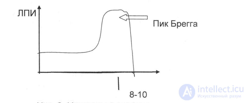
Fig. 1. Protons with an energy of 160-180 MeV
An electron beam with an energy of up to 5 MeV is used in the treatment of superficial malignant neoplasms, and with an energy of 20 to 50 MeV - for more deeply located ones. Modern accelerators make it possible to smoothly adjust the energy of the electron beam and thereby create the required dose at any depth.
Sources of electrons are ELA (electron linear accelerators), betatrons, and microtrons. The maximum absorbed dose is located at the depth of the effective electron range (the effective range equals 1/3 of the maximum energy), i.e., 7-10 cm from the irradiated body surface. The dose value drops rapidly with depth. It is mainly used for repeat RT or for treating tumors located near critical organs.
- Gamma-therapy.
Gamma radiation arises during radioactive decay. The transition of the nucleus from an excited to the ground state is accompanied by the emission of a gamma quantum with energies from 10 keV to 5 MeV. The main therapeutic sources of gamma radiation are gamma units (guns).
A radionuclide is used as the radiation source (until recently - cesium-137, currently - cobalt-60). Requirements for radionuclides for gamma units:
1. The physical half-life must be long: S cesium-137 - 33 years; S cobalt-60 - 5.3 years.
2. The energy of gamma rays must be sufficient (1 MeV or more): S the energy of cesium gamma rays - 0.66-0.75 MeV; S the energy of cobalt gamma rays - 1.17-1.33 MeV.
3. The preparation must have a relatively high specific activity (radionuclide activity per unit volume). The higher the specific activity, the smaller the size of the radiation source. Since the specific activity of cobalt is greater than that of cesium, it is more convenient to use it in the clinic (currently the dimensions of a cobalt pellet are 1.6 x 1.6 cm).
The maximum absorbed dose during gamma-therapy is
продолжение следует...
Часть 1 6. Devices and systems for exposure to X-ray and radioisotope radiation.
Часть 2 Contact methods of irradiation - 6. Devices and systems for

Comments