
Lecture
At Vanderbilt University, a microcomputer system was developed that makes it possible to design, select and dose psychomotor tests and monitor their performance. At the same time, an assessment is made of the residual blood nitrogen level, since patients with a high residual nitrogen content show a worse response to the test than patients with a normal nitrogen content. The microcomputer performs four types of tests: reaction time (two kinds), attention, and short-term memory.
The system uses an Intelligent Systems Corp. 8080 microprocessor. The Model 8001 terminal is used to store the program and to display various images on the screen. To respond, the subject must press a button. Apart from the terminal, the system uses only one peripheral device — a key.
The microprocessor offers the following tests. One of four possible symbols must be selected in a random sequence. If the selected symbol matches the symbol shown on the display as specified, the patient presses the key. The reaction time is measured by the microcomputer. To assess attention (scanning ability), the number of times the subject recognized a given symbol during its threefold presentation in a row of 16 symbols is taken into account. The short-term memory test consists of determining whether the symbol shown on the display had been presented before. The program generates random numbers for each test, reproduces an individual test, and calculates the average reaction time for subsequent evaluation. The program is loaded directly from another computer.
Life-support system for the elderly
With age, short-term memory is often weakened or lost. Because of this, patients who need to restrict their diet and take medication themselves cannot manage without outside help. To create a reminder device with displayed information, a 6502 microcomputer with 4 Kbyte of RAM is used. After pressing a button, the patient sees on their television screen the daily schedule and the time for performing the necessary procedures, for example: insulin injection, breakfast menu, four capsules of medicine, measuring and recording blood pressure, a nurse's visit, a dentist's visit, a meal from a mobile trolley. If the patient forgets to carry out the procedure at the specified time or to cancel it, an alarm signal will sound. New information is periodically entered by the person looking after the patient.
Registration of ultra-slow physiological processes using Omegametry.
The study of electrical processes associated with the vital activity of the organism began at the same time as the discovery of electrochemical processes. The first publication on the presence of currents in the central nervous system was made in 1849. In 1912, P.Yu. Kaufman revealed a connection between the electrical potentials of the brain and the ‘‘internal activity of the brain’’ and their dependence on changes in brain metabolism and the effects of external stimuli.
The problem of choosing methods for assessing functional states, the compensatory-adaptive capabilities of the main regulatory systems, and stress resistance under changing conditions of the external and internal environment of the organism has not yet been fully solved. Alongside such methods as EEG, ECG, the Voll method and others, there is the method of omegametry, which makes it possible to diagnose the body's regulatory systems fairly quickly and easily.
Studies of the spontaneous dynamics of ultra-slow physiological processes, recorded in the brain and in non-cerebral structures, lying in the frequency band from 0 to 0.5 Hz, make it possible to assess the level of the functional state of various organs and systems.
In experimental physiological-biochemical and pharmacological studies at the cellular, tissue and structural level, direct evidence has been obtained of the connection between the dynamics of various types of ultra-slow physiological processes (USPP) of the brain, liver, kidneys, lungs and muscles with oxidation-reduction processes (including the processes of glycolysis, gluconeogenesis and oxidative phosphorylation), energy and mediator metabolism, RNA-synthesizing activity of cells, and the intensity of neurosecretory and hormonal activity of tissues and organs; a dependence has been revealed of the dynamics of USPP of the brain and glandular organs (skin, salivary glands, glands of the tongue, pharynx, stomach) on their secretory activity, as well as on exogenous influences (including oxygen deficiency, temperature, mechanical and electrical stimuli); the influence of the blood supply of the organ and tissue on the dynamics of USPP has been established.
A similarity has been found between the temporal characteristics of the ultra-slow physiological processes of the brain, autonomic reactions, psychological indicators of attention, memory, emotions, and other adaptive behavioral reactions .
Currently, existing omegametry techniques make it possible to determine the level of wakefulness, characterizing the body's ability to learn and react to external stimuli: light and acoustic vibrations of various frequencies, temperature factors and others.
Omegametry is a theoretically substantiated method for solving problems of rapid diagnosis of functional states, stress resistance, the compensatory-adaptive capabilities of the main regulatory systems, and compensation reserves in healthy and sick individuals.
It has been established that in response to a certain stimulus, the central nervous system reaches its operating mode within tenths of a second.
Measurements are performed using two silver-chloride reference electrodes, Ers-10101 and Ers-10102. The KCl concentration is 4.2 mol/dm3. One electrode is placed in the vertex area (crown of the head), the point being located at the intersection of the head's midline and the line connecting the ears. The second electrode is applied to the back of the hand between the thumb and index finger (the thenar of the hand), to the left or right hand depending on whether the subject is left-handed or right-handed. The electrodes are connected to a voltmeter, from which readings are taken. The subject sat in a relaxed state with eyes closed (the initial value of the omega-potential was measured) until the omega-potential value stopped changing, then, from a standing position, performed 5 squats and sat down again. During the first minute, the omega-potential was measured every 10 seconds, thereafter every 30 seconds. The obtained data were then analyzed according to the following scheme: 1) the first 30 sec. after the functional load characterizes the state of the neuroreflex and autonomic mechanisms regulating the respiratory and cardiovascular systems that ensure the delivery of oxygen to the tissues. 2) The following 30 sec.-1.5 min. reflects the state of the mechanisms ensuring the processes of oxygen consumption and utilization in the tissues, primarily in brain tissue (tissue respiration processes). 3) The following 1.5-3.5 min. after the functional load characterizes the state of the neurohumoral mechanisms ensuring the coordinated interaction of the visceral systems performing detoxification (gastrointestinal tract, liver, kidneys). 4) The following 3.5-6 min. after the functional load characterizes the state of the mechanisms of neurohumoral regulation ensuring the coordinated interaction of internal organs under any stress exposure on the body. A conclusion about the degree of excitability of the individual is drawn from the initial value of the omega-potential.
When recording the background value, measurement of the omega-potential begins from the moment the electrodes are connected to the subject; the study is carried out over 10 minutes, and the omega-potential value is measured every 10 seconds.
The background value of the omega-potential after reaching a plateau reflects the level of wakefulness and the nonspecific resistance of the organism to stress exposures. The characteristic parameters are: the sign and magnitude of the omega-potential (mV) after reaching the plateau; the direction, intensity and character of the changes in the omega-potential before reaching the plateau; the time to reach the plateau (min); the presence and character (regular, aperiodic) of ultra-slow potential oscillations, their amplitude and dominant period.
Typology and physiological significance of evoked changes in ultra-slow processes
As a result of generalizing the materials accumulated to date on the features of the evoked dynamics of the omega-potential in response to a single functional load in healthy individuals and patients of different age groups, the most common types of evoked omegagrams have been identified. Their diagnostic significance has been analyzed with greater or lesser completeness [8,11,12,13]. The type of evoked omegagram is formed from the combination of the features of omega-potential dynamics successively in each of the phases described above.
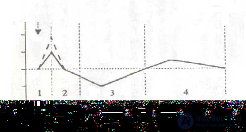
Figure 1 - The first type of evoked omegagram
The first type of evoked omegagram is characterized by the occurrence of a negative deviation of the potential from the initial values in the first 30 sec. after the load (phase 1).
With an optimal state and balance of the mechanisms of neuroreflex and autonomic regulation of the bronchopulmonary apparatus and the cardiovascular system, which ensure the processes of oxygen delivery to the tissues, the deviation of the omega-potential in the first 30 sec. after the load reaches 4-12 mV.
With compensatory hyperactivity of the mechanisms of neuroreflex and autonomic regulation of the bronchopulmonary and cardiovascular systems (with a predominance of sympathetic influences of the autonomic nervous system under these conditions), the deviation of the omega-potential in the first 30 sec. reaches 13-18 mV or more.
Then, in the following 30 sec.-1 min. (2nd phase), a decrease in the omega-potential to the initial or somewhat below the initial (background) values of this parameter is noted. This kind of direction of omega-potential dynamics in the 2nd phase characterizes the optimal state of the mechanisms regulating the processes of oxygen utilization by the tissues (tissue respiration), including at the level of the CNS.
The third phase of omega-potential dynamics (in the interval from 1.5-2 to 3.5-4 min.) is characterized by the occurrence of a trough-shaped positive wave, the leading edge of which is represented by a smooth decrease in the omega-potential from the initial values over 1.5-2 min., with a subsequent return to the initial level by the 4th min. after the functional load. This kind of omega-potential dynamics is observed with a balance of the mechanisms of neurohumoral regulation of all links of the detoxification system at the level of the gastrointestinal tract, liver, and kidneys.
The fourth phase of the evoked omegagram (in the interval from 4 to 7 min. after the load) is characterized by the occurrence of a negative wave with a peak latency of 5 min. This kind of evoked change in the omega-potential reflects the optimal state of the mechanisms of neurohumoral regulation of the hypothalamic-pituitary-adrenal system, which ensures the coordinated interaction of internal organs in response to any kind of signal from outside.
Thus, type I of the evoked omegagram characterizes a mixed or sympathetic-adrenal orientation of the autonomic support of the reactions of the external respiration system and cardiovascular reactions, an optimal state of the mechanisms of neuroreflex and autonomic regulation of hemodynamics, tissue respiration processes, neurohumoral regulation of the body's detoxification systems, and the functions of the hypothalamic-pituitary-adrenal system, which form an adequate response to stress exposure.
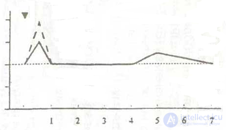
Figure 2 - Subtype of the first type
A subtype of the first type of evoked omegagram has been identified (1a), distinguished by the absence of omega-potential dynamics in the interval from 1.5 to 3.5-5 min. after the load, which indicates arreactivity of the mechanisms of neurohumoral regulation of the detoxification functions of the visceral systems (GI tract, liver, kidneys).
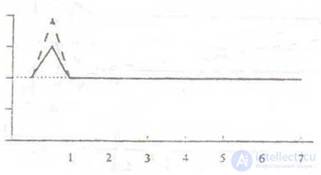
Figure 3 - The second type of evoked omegagram
The second type of evoked omegagram is characterized by an increase in the omega-potential to 10-39 mV from the initial values in the first 30 sec after the load, followed by a decrease to the initial values within 30 sec.-1 min, with no changes in the 3rd and 4th phases. This type of omegagram reflects: a) high reactivity of the mechanisms of neuroreflex and autonomic regulation of the bronchopulmonary and cardiovascular systems, with a pronounced predominance of sympathetic tone of the autonomic nervous system; b) arreactivity of the neuroreflex mechanisms ensuring the detoxification functions of the GI tract, liver, and kidneys; c) arreactivity of the neurohumoral mechanisms of regulation of the hypothalamic-pituitary-adrenal system, which ensures the organization of the body's adaptive response to stress exposure.
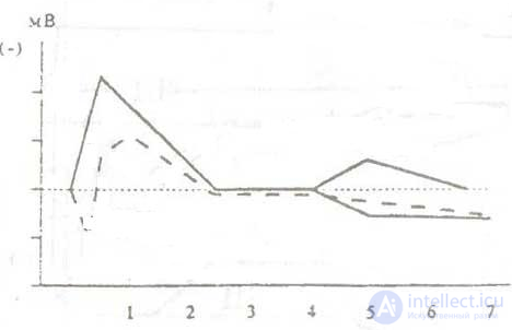
Figure 4 – The third type of evoked omegagram
The third type of evoked omegagram is characterized by a sharply pronounced negativation of the omega-potential in the first 30 sec after the load (up to 20-30 mV and higher), followed by the maintenance of these high values (or an insignificant decrease) for 1-1.5 min., a further decrease to the initial values by the 3rd-4th minute after the load (with the occurrence of a characteristic negative, hump-shaped wave), and a continued decrease by the 7th minute. This type of omegagram characterizes: a) a pronounced hyperactivity of the mechanisms of neuroreflex and autonomic regulation of the bronchopulmonary and cardiovascular systems, which ensure the delivery of oxygen to the tissues; b) a disruption of the neurohumoral and enzymatic mechanisms ensuring the processes of tissue respiration and, as a result, autointoxication by underoxidized metabolic products; c) functional insufficiency of the neurohumoral mechanisms of regulation of the hypothalamic-pituitary-adrenal system.
The third type of evoked omegagram correlates with the clinical and laboratory manifestations of the hyperdynamic type of blood circulation, a tendency toward arterial hypertension, manifestations of acidosis, activation of the blood aggregate state regulation system, and a tendency toward decreased nonspecific resistance of the organism. Essentially, one of the varieties of type III omegagram is type IIIa, the main distinction of which lies in the occurrence of the first short-latency (5-15 sec.) positive wave with an amplitude of 6-15 mV, followed by negativization of the potential and the formation of the same "hump-shaped" negative wave as in type IV, with further stabilization at background values or a slight decrease by the 7th minute.
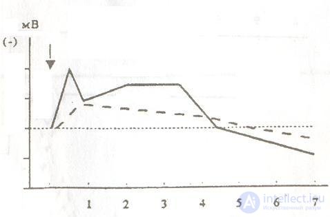
Figure 5 - Fourth type of evoked omegagram
The fourth type of evoked omegagram differs from the third by the prolongation of the duration of the high-amplitude (up to 20-30 mV) negative wave in the interval from 30 sec.-1 min. to the 4th-5th minute after the load, followed, as a rule, by a significant decrease in the omega-potential (below background values) by the 7th minute after the load. Variations in the amplitude of the prolonged negative wave are possible. This type of omegagram is characterized by: a) pronounced hyperactivity of the neuroreflex and autonomic regulation mechanisms of the bronchopulmonary and cardiovascular systems that ensure the delivery of oxygen to the tissues; b) the presence of autointoxication due to disruption of the mechanisms ensuring tissue respiration, as well as insufficiency of the neurohumoral support of the detoxification functions of the digestive and urinary systems; c) functional insufficiency of the hypothalamic-pituitary-adrenal system.
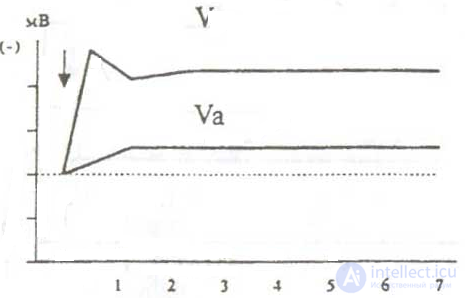
Figure 6 - Fifth type of evoked omegagram
The fifth type of evoked omegagram is characterized by a sharp rise in the omega-potential (up to 30 mV and above) in the first 10-30 sec. after the load and the maintenance of these high values until the end of the study. This type of change in the omega-potential reflects pronounced psychoemotional tension, the presence of psychoemotional disorders of the hypersthenic type with a tendency toward the occurrence of affective states.
Changes in the omega-potential that are similar in direction but substantially differ in intensity have been distinguished as the Va subtype. This subtype of evoked omegagram is characterized by a small rise in the omega-potential in the first 30 sec. after the load (within 10 mV) and the subsequent maintenance of these values or their gradual smooth change (without reaching the initial values) over the following 7 min. after the load. This type of change in the omega-potential correlated with the clinical manifestations of encephalopathy, intoxication, and changes in the acid-base balance
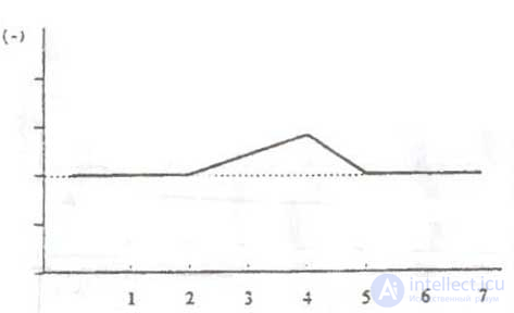
Figure 7 - Sixth type of evoked omegagram
The sixth type of evoked omegagram is characterized by the absence of dynamics of the omega-potential in the first 30 sec.-1 min. after the load, the occurrence of a trough-shaped negative wave in the interval from 1.0-1.5 min. to 5 min. with further attainment of the initial values by the 6th-7th minute after the load. As comparative clinical and physiological studies have shown, the absence of dynamics of the omega-potential in the first 30 sec.-1.5 min. after the load is an indicator of the areactivity of the neuroreflex and autonomic mechanisms that ensure the delivery of oxygen to the tissues and the mechanisms of tissue respiration. The trough-shaped negative wave in the interval from 1.5 to 5 min. after the load indicates the presence of autointoxication by underoxidized metabolic products due to insufficient hemodynamic compensation of metabolic lactic acidosis and the oxygen debt. In contrast to type VI, the distinguished subtype VIa shows an intensification of the above-described phenomena of autointoxication due to functional insufficiency of the hypothalamic-pituitary-adrenal system.
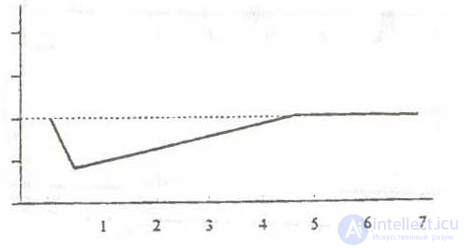
Figure 8 - Seventh type of evoked omegagram
The seventh type of evoked omegagram is characterized by a decrease in the initial values of the omega-potential in the first 30 sec. after the functional load, followed by a smooth return of its values to the initial background by the 4th minute after the load and the occurrence of a negative monophasic wave with peak latency at the 5th minute after the load.
This type of evoked omegagram correlated with the insufficient oxygen-transport and hemodynamic compensation, revealed under these conditions, of the deepening metabolic acidosis and mixed hypoxia. In contrast to type VII of the evoked omegagram, in the VIIa subtype we distinguished, there was an absence of changes in the omega-potential in the interval from the 4th to the 7th minute after the load, which characterized the areactivity of the neurohumoral regulatory mechanisms of the hypothalamic-pituitary-adrenal system, aggravating the above-described phenomena of metabolic acidosis and mixed hypoxia under functional insufficiency of the neuroreflex regulatory mechanisms of the cardiovascular and respiratory systems that ensure the delivery of oxygen to the tissues and tissue respiration.
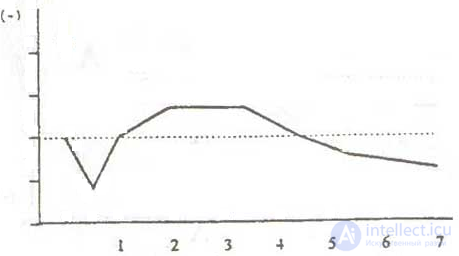
Figure 9 - Eighth type of evoked omegagram
The eighth type of evoked omegagram is characterized by a decrease in the omega-potential from the initial values in the first 30 sec. after the load, a return to the initial background by the 1st minute after the load, the occurrence of a trough-shaped negative wave in the interval from 1-1.5 min. to 3-4 min. after the load, followed by a decrease in the omega-potential by the 7th minute significantly below the initial values. This type of omegagram correlated with the clinical and laboratory manifestations of encephalopathy, mixed hypoxia, disturbances of the acid-base balance, and a decrease in nonspecific resistance.
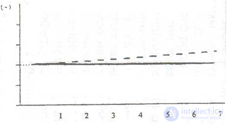
Figure 10 – Ninth type of evoked omegagram
The ninth type of evoked omegagram is characterized by the absence of changes in the omega-potential in response to the functional load. This type of omegagram correlates with the clinical and laboratory manifestations of areactivity of the sympathoadrenal and pituitary-cortical system, and is found in individuals with pronounced encephalopathy, multiple organ failure, and disorders of the cardiovascular, respiratory, and detoxifying systems of the body. A prognostically unfavorable sign is the detection of the IX type of evoked omegagram at very low background values of the omega-potential. The IX type of omegagram at low background values of the omega-potential (2-15 mV) was found in patients with peritonitis who had a fatal outcome after surgical intervention.
The detection of the same type of evoked omegagram against the background of average values of the omega-potential (24-40 mV) was a prognostically favorable sign for the course of the postoperative period in patients with peritonitis.
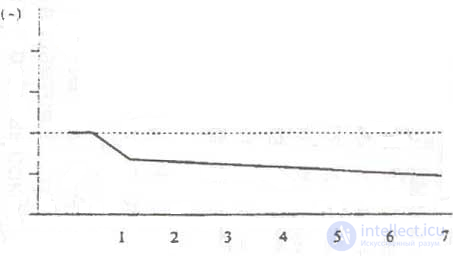
Figure 11 - Tenth type of evoked omegagram
The tenth type of evoked omegagram is characterized by the absence of changes in the omega-potential in the first 30 sec. after the load, followed by an abrupt decrease over 30 sec. and the maintenance of the decreased values for 7 min. or more.
This type of evoked omegagram correlates with the clinical and laboratory manifestations of functional insufficiency of the pituitary-adrenocortical system, which was reflected in pronounced lymphopenia and a tendency toward leukopenia.
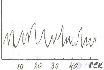
Figure 12 - Eleventh type of evoked omegagram
The eleventh type of evoked omegagram was characterized by the appearance (persistence or intensification) of regular, deformed, sharpened ultra-slow waves with a tendency toward rhythmicity in the deca-second and second range (tau and zeta waves). This type of omegagram reflects the presence of varying degrees of metabolic tension against the background of encephalopathy, and correlates with the clinical and laboratory manifestations of the activation of compensatory mechanisms aimed at eliminating metabolic acidosis.
The proposed typology of spontaneous and evoked omegagrams recorded from the vertex-thenar (or forehead-thenar) leads of the hands is the result of a synthesis of all known and methodically correctly performed studies in this field. It is evident that the types of omegagrams have no nosological affiliation, but reflect the state of the neuroreflex, autonomic and neurohumoral mechanisms regulating the levels of activation and adaptive reactions of the organism, and make it possible to judge the adaptive reserves and adaptive capabilities of the main regulatory systems that ensure the interaction of the organism with environmental factors [15].
Systems for psychological research.
Psychology is based on physiology, and primarily on the physiological processes occurring in the brain.
Studies conducted (in the USA) in recent years have shown that the teenage brain demonstrates less activity in the area associated with motivation. It is possible that teenagers' love of dangerous activities is related precisely to the underdevelopment of this key area of the brain. Teenagers are more willing than adults to take risks – they use drugs and alcohol more often and engage in sexual excesses – but the reasons for this are the subject of heated debate. Now researchers from the National Institute on Alcohol Abuse and Alcoholism (USA) have found differences in the brain chemistry of teenagers and adults – in the part that is associated with the assessment of risks and rewards. In teenagers, this part of the brain, the ventral striatum, is insufficiently active, showed the author of the study, James Bjork. He suggested that teenagers get involved in risky adventures in order to achieve a normal level of stimulation of this part of the brain.
Bjork and his colleagues scanned the brains of 12 teenagers and 12 adults while they played a special gambling game, in which they received different financial rewards for achieving the goal. Although, when questioned, both teenagers and adults assessed the value of the rewards received in the same way, and achieved the goal of the game with the same frequency, the brain scan showed that this organ works differently in them. The right ventral striatum of the teenagers demonstrated much less activity than that of the adults.
It appears that the brain circuit of motivation and reward in teenagers is underexpressed, so they require extreme stimuli to achieve the same level of brain activity as adults. The difference in activity may become even more pronounced if the reward is not immediate – perhaps that is precisely why it is so difficult for teenagers to achieve long-term goals". – commented the researcher.
Motivation.
There are several different styles of motivation. They are easy to identify by looking at the everyday ritual of getting out of bed. When the alarm clock rings in the morning, what happens inside you? Although, it would be more correct to ask: What do you do inside yourself? Perhaps your inner voice tells you: "I need to get up, otherwise I'll be late, and I'll have big problems". Your brain begins to paint pictures of you being late for work, and images of the problems that could arise from this. And when these pictures, seen through the eyes of your imagination, become big enough, you say to yourself: "Okay, okay, I'm getting up". Finally, you are motivated enough to get up. People who mobilize themselves to take certain actions by creating a scenario of what they would like to avoid act approximately as described above.
+Now let us describe a different type of motivation. You open your eyes and begin to think about what you could do today. Vivid pictures of attractive possibilities appearing in your head pull you out of bed like a giant magnet. The only question that arises for you is: "What should I do first?" The question of whether to get up at all does not even come to mind! Surely each of you, dear readers, can recall more than one morning when you woke up and thought about the wonderful things you had ahead of you that day. You thought about how you would engage in something pleasant for you, with great joy, anticipation of success and self-confidence. You saw yourself performing these actions and moving toward the reward for accomplishing them. You realized that this day connects you with the next, bringing you closer to what you truly strive for. Psychology has long known these two described types of motivation. One of them is called - avoidance of failure, and the other - the motive of achieving success. These types of motivation act in different ways, in different directions and with different results. All people, to a certain degree, use both, but each of us tends to give preference to one of these directions. For example, someone who cannot get out of bed until they clearly picture their boss threatening to fire them will most likely be guided by the motivation of avoiding failure in many other situations. When choosing friends, he will more likely focus attention on those people who will not "pressure" him. He will not decide to change jobs until he realizes that he can no longer stand the one he is currently working at. With a predominance of the motivation of achieving success, a person who wakes up early in the morning cannot wait to jump out of bed and start realizing their dreams. Most often, in this case, the individual will apply this type of motivation in other cases as well. For example, when deciding what moment will be the best one to take a break from work - to chat with a colleague or reward themselves with a cup of tea for finishing something. He will most likely change jobs for a possibly better one, if such an opportunity arises.
At first glance, when comparing the two types of motivation, it may seem that the strategy of achieving success is more attractive. As practice shows, both types of motivation have their positive and negative sides. For example, some people are so seriously focused on achieving their goal that they do not even think about the problems that may arise in their path. Such thinking is typical of some young novice entrepreneurs. The other extreme - people whose motivation to avoid failure is so strong that they are too frightened to make any attempt. Since both strategies are important, we can use them to motivate other people. This knowledge is especially useful for directors and managers. By observing people's behavior, it is easy to determine that someone is mobilized to a greater degree by rewards and praise. These people, if they know they will be rewarded, will work very hard. With respect to such people, a successful director will apply incentives for achieving the goal: setting goals and giving bonuses. For those who prefer the strategy of avoiding failure, the same bonuses will not have much significance. As a manager, you may be surprised by such a situation: "I offer them any rewards, and they still haven't lifted a finger". If you express your displeasure in a calm, measured tone, warning of possible dismissal, they suddenly, out of nowhere, start working like crazy.
In conclusion, I will mention one more area of application of knowledge about the two types of motivation. When describing, for example, new goals for an organization, you will most likely immediately receive the approval of those in whom the motivation of achieving success predominates. Soon, members of the team with the motivation of avoiding failure will also join the conversation, saying that nothing will come of it, giving various arguments why. It is very important that they be heard. And what they say can show you the pitfalls of the situation.
To clarify
Various psychological tests are used. Let us consider one of them using the example given below.
Draw a house: the sheet positioned lengthwise. Questions about the drawing:
1. What is it made of?
2. How old is it?
3. Who built it?
4. How many rooms does it have?
5. Which room is the most beautiful and why?
6. Which room is the worst and why?
7. Is the house sturdy or dilapidated?
8. Who lives in this house? Are they cheerful or sad?
9. Why exactly did you draw this house?
Additional questions about the drawing: why is this or that here, especially extra details.
Draw some tree, other than a fir tree; the sheet positioned crosswise. It is better to use a square sheet for this. Questions:
1. What kind of tree is this,
2. How old is it?
3. How does it grow: in the wild or in a garden?
4. Was it planted, or did it grow by itself?
5. How many years will it live?
6. Is the climate good or bad?
7. Is the tree sick or healthy?
8. Have you seen this tree before? Why did you decide to draw it in particular.
* Drawing of a person; the sheet positioned crosswise.
1. How old is he?
2. What is his name?
3. Is he healthy or not?
After this, decoding is performed in accordance with a special program.
Decoding of various drawings is also used. For example, the following:

The Wife and the Mother-in-law
Most people, at first glance, see a young woman standing at a half-turn. But if you look closer? Imagine that the girl's necklace is her mouth, and her whole profile is a nose, what do we see...

The Husband and the Father
J.Botwinick, "My husband and my father-in-law" Approximately the same as in the previous one. A young man in half-turn, or an elderly man, in profile.

The Indian - The Eskimo
What do you see? The head of an Indian, proudly looking at the sun? Or, perhaps, an Eskimo in a hood, turned with his back...

The Duck – The Rabbit

Mother, Father and Daughter.
On the left - the daughter, on the right the mustached father, and in the middle - the mother. Drawing - G.H.Fisher
|
|
|
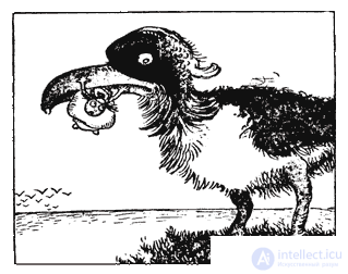
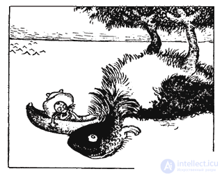
The Bird - The Tree
+This is a picture from the series of reversible images by Gustave Verbeek. If you look at it normally you will see a bird. But if you turn it over - the bird forms an island, its legs - trees.

Comments