
Lecture
Это окончание невероятной информации про .
...
use is in fact mistaken: modern techniques for identifying substances do not use the colors of the mixture's components. One of the phases (liquid or solid) is stationary, while the other (liquid or gaseous) is mobile. If a liquid phase is used as the stationary one, the process is called partition. If a solid phase is used as the stationary one, the process is called adsorption.
To separate the components of a mixture, chromatology makes use of the difference in the speed of their movement in the mobile phase, caused by the interaction of these substances with the stationary phase.. Four possible combinations of mobile and stationary phases are used in chromatographic methods.
From the standpoint of clinical laboratory applications, these methods are used primarily to detect complex substances such as drugs and hormones. For example, gas-liquid chromatography (GLC) and thin-layer chromatography (TLC) are convenient for determining which particular drug (or drugs) was used in a case of overdose. Having this information is vitally necessary for the physician, who must choose the appropriate therapeutic procedures..
The main elements of a GLC are shown in Fig. 11.7. Before being introduced into the GLC, the sample obtained from the patient is usually subjected to a preliminary cleanup procedure, the extent of which depends on the analysis being performed. The main subsystems of the chromatograph perform the following functions: Injector. The injector is designed to introduce 1—5 mL of the sample solution (usually in a volatile organic solvent) into the GLC. The temperature in the injector is such that the solvent and the sample evaporate instantly.
Carrier gas. An inert carrier gas (usually N2 or He) is the mobile phase of the chromatograph. It carries the vaporized sample and gaseous solvent down the column.
Column. The column is usually 1 m long and less than 7 mm in diameter. It is packed with a solid supporting matrix (for example, diatomaceous earth). A liquid phase is applied to the solid matrix. The small size of the solid granules ensures separation of the components. The column is placed in a thermostat whose temperature is carefully controlled. A temperature programmer gradually increases the column temperature in a sequence chosen to achieve maximum efficiency in isolating the particular substance being analyzed.
Detector. The detector is located at the end of the column. Its function is to produce an electrical signal proportional to the amount of substance in the outgoing gas stream. There are a number of types of detectors designed for use with different types of samples. These include ionization detectors, thermal-conductivity detectors, and electron-capture detectors. Ionization detectors are the ones most often used for clinical laboratory applications (Littlewood, 1970).
All detectors are sensitive to entire classes of substances rather than to any one particular analyte. Thus, to determine the types and amounts of individual components present in a sample, both the concentrations of the detected substances and the time periods after which these concentrations are recorded are used (i.e., a graph of concentration versus time). The detector's electrical output is connected to a recorder.
Recorder. On the recorder, time is plotted on the x axis, and the intensity of the detector's output signal — on the y axis. Thus, the chromatogram shows both the amounts of the components present (the areas of the corresponding peaks) and the retention times at which these substances elute from the column. Based on this information, the components present can be identified by their retention times or, preferably, by comparison with chromatograms obtained from GLC analysis of substances of known structure.
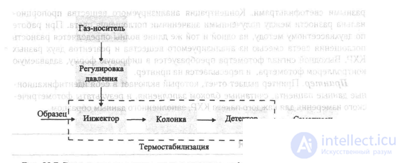
Fig. 11.7 Block diagram of a gas-liquid chromatograph (GLC).
Fig. 11.8 shows a chromatogram obtained from analyzing a blood sample for the content of known anticonvulsant drugs — phenobarbital and phenytoin. A known amount of heptabarbital was added to the sample as an internal standard. To calculate the levels of phenobarbital and phenytoin, the areas of the peaks corresponding to these drugs were compared with the area of the heptabarbital peak. Gas-liquid chromatography offers a number of important advantages in the analysis of complex compounds: speed, the ability to work with small sample amounts, and high sensitivity. Most instruments can analyze clinically significant substances in less than 1 h, and often even in less than 15 min, with only about 1 mL of sample needed for the analysis. The sensitivity of the instrument depends on the type of detector used. High-quality instruments are capable of analyzing 1 ng of substance.
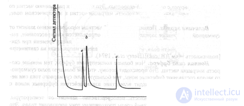
Fig. 11.8 Example of a GLC chromatogram for analysis of blood levels of phenobarbital (peak a) and phenytoin (peak c). Peak b corresponds to the heptabarbital level (internal standard).
HEMATOLOGY
BASIC CONCEPTS
Blood consists of formed elements, dissolved substances, and water. This section describes only the instruments that measure the characteristics of the formed elements: red blood cells (RBC), white blood cells (WBC), and platelets (PLT). The main function of RBCs is to carry oxygen from the lungs to various organs and to carry carbon dioxide from these organs to the lungs for its removal from the body. The main function of WBCs is to participate in defending the body against infections. Five types of WBCs are normally found in peripheral blood. In decreasing order of content in the blood of an adult, these are neutrophils, lymphocytes, monocytes, eosinophils, and basophils. In disease states, the total counts and proportions of these types of WBCs can change; immature and malignantly transformed types of WBCs can also appear. Platelets plug small breaks in the walls of blood vessels and also take part in the blood clotting mechanism.
The main measure of the blood's formed elements is the number of elements of each type per microliter (uL) of blood. The normal range of RBC content for an adult man is 4.6—6.2 x 10 /uL, and for an adult woman 4.2—5A x 106/uL. The normal ranges of WBC and platelet content are the same for men and women. The normal range of WBC content is 4500— 11000/uL, and the normal range of platelet content is 150000—40000/uL. Hematocrit (HCT) is the ratio of the volume of all the blood's formed elements to the total volume of the blood sample. Hematocrit is expressed as a percentage, and its normal range for an adult man is 40—54%, and for an adult woman 35-47%. Hemoglobin (Hb) is a conjugated protein located inside the RBCs. This protein transports most of the O2 and part of the CO2 carried by the blood. Its content is expressed in grams per deciliter. The normal range for an adult man is 13.5—18 g/dL, and for an adult woman this range is 12—16 g/dL.
+A second group of measurements is performed to assess RBC cell volumes and Hb concentration. These measurements include determining the mean cell vol-
Часть 1 16 Equipment for Analyses
Часть 2 Gas-liquid chromatographs - 16 Equipment for Analyses

Comments