
Lecture
The set of processes associated with the consumption and assimilation by the body of substances that make up food is called digestion. Food is a source of energy and structural (plastic) materials for the body. It enters from the external environment in the form of raw food materials or products that have undergone technological processing. Raw food materials and food products include a multitude of nutritional substances, as well as non-nutritional compounds and foreign impurities.
The selection and extraction from food of the substances necessary for the body and their conversion into a form that will be available for assimilation by tissues is carried out by the digestive system. As a result of its activity, food undergoes digestion, i.e., physical, physicochemical, and chemical transformations that promote the formation of monomers from polymers, which are absorbed and assimilated by the body. Physical changes in food consist of its grinding, mixing, the formation of suspensions and emulsions, and partial dissolution. Chemical changes consist of the breakdown
of proteins, fats, and carbohydrates into smaller compounds.
A distinction is made between cavity (luminal) digestion and membrane digestion: cavity digestion → membrane digestion → absorption
Cavity digestion is digestion that takes place in the digestive cavities of the body – the oral, gastric, and intestinal cavities. This type of digestion provides intensive initial digestion.
Membrane (parietal) digestion is carried out with the help of enzymes concentrated on the microvilli in the small intestine. Membrane digestion carries out the intermediate and final stages of hydrolysis of food components, as well as the transition from the final stages of digestion to the initial stages of absorption.
What it is: the earliest stage of digestion, which begins even before food enters the mouth.
Mechanism:
Occurs at the sight, smell, taste, or even thought of food.
The central nervous system is activated (mainly the vagus nerve).
Signals stimulate the salivary glands and the stomach to secrete their secretions.
Significance:
Prepares the body for the intake of food.
Enhances the secretion of saliva, gastric juice, and sometimes even pancreatic enzymes.
Ensures more efficient digestion.
Mechanical processing: chewing grinds the food.
Mixing with saliva: the enzyme amylase begins the breakdown of starch.
Formation of a food bolus: convenient for swallowing.
Duration: 15–20 seconds.
Chemical processing: gastric juice (hydrochloric acid, pepsin) breaks down proteins.
Motor function: mixing of food, converting it into chyme.
Duration: 1–10 hours.
Enzymatic breakdown: pancreatic enzymes and bile break down proteins, fats, and carbohydrates.
Absorption: nutrients enter the blood and lymph.
Duration: about 6 hours.
Absorption of water and electrolytes.
Formation of fecal matter.
Excretion: elimination of undigested remnants.
Duration: 24–72 hours.
Comparative table of the phases of digestion
| Phase | Main processes | Transit time | Regulation | Key enzymes/media |
|---|---|---|---|---|
| Cephalic | Nerve signals at the sight/smell of food, stimulation of secretion | CNS, vagus nerve | ||
| Oral | Chewing, saliva, amylase | 15–20 seconds | Local reflexes | Saliva, amylase |
| Gastric | Protein digestion, secretion of HCl and pepsin | 1–10 hours | Gastrin, nerve impulses | Pepsin, HCl |
| Small intestine | Enzymatic breakdown, absorption | ~6 hours | Secretin, cholecystokinin | Pancreatic enzymes, bile |
| Large intestine | Absorption of water, formation of fecal matter | 24–72 hours | Microflora, local reflexes | Intestinal microflora |
Important points
The regulation of digestion is carried out by the nervous system and hormones (for example, gastrin, secretin).
The rate at which food passes through depends on its composition: proteins are retained longer than carbohydrates.
The intestinal microflora plays a key role in the breakdown of fiber and the synthesis of vitamins.
The digestive system includes the digestive canal, or gastrointestinal tract (GIT), the pancreas, and the liver (Figure 36).
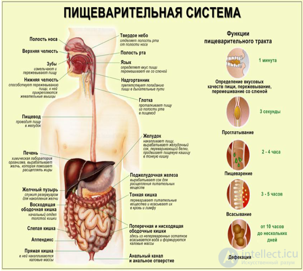
Figure 36 – Structure of the gastrointestinal tract
The digestive canal consists of separate cavities and passes through the entire body; it begins with the oral cavity and ends with the opening of the rectum –
the anus.
The gastrointestinal tract can be conditionally divided into three parts:
1. Oral cavity – passes into the pharynx, from which food enters the esophagus.
2. Stomach.
3. The intestine consists of two sections:
- small intestine – consists of the duodenum, jejunum, ileum); the length of the small intestine is 5-7 m, diameter 3.0-3.5 cm;
- large intestine – consists of the cecum with the appendix, the colon (the ascending section of the large intestine, the transverse section of the large intestine, the descending section of the large intestine), the sigmoid colon, the rectum, the anus; the length of the large intestine is 1.5-2 m, diameter in the upper sections 7 cm, in the lower sections about 4 cm.
The process of breaking down natural biopolymers (proteins, fats, carbohydrates) is carried out in the body by enzymatic hydrolysis with the help of digestive enzymes belonging to the 3rd class of enzymes – hydrolases. Only macronutrients are depolymerized (broken down to monomers). Three groups of enzymes of the hydrolase class participate in depolymerization:
- proteases (enzymes that break down proteins);
- lipases (enzymes that break down fats);
- amylases (enzymes that break down carbohydrates).
Enzymes are formed in special secretory cells of the digestive glands and enter the digestive tract together with saliva and
digestive juices – gastric, pancreatic, and intestinal, the volume of secretion of which is about 7 liters per day.
For effective digestion, a set of enzymes providing comprehensive action is necessary. They are produced by the digestive glands. A list
of the digestive enzymes of the GIT is presented in Table 10.
Table 10 – Digestive enzymes of the GIT
Digestive
enzymes
Optimal
pH value Substrate
Enzymes that digest proteins
pepsin 1.0-1.5 most proteins
of a globular nature
gastricsin 2.0-3.0 most proteins
of a globular nature
trypsin 8.0 most proteins
of a globular nature
chymotrypsin 8.0 most proteins
of a globular nature
aminopeptidase 8.0 peptides from the N-terminal
amino acid residue
carboxypeptidase 8.0 peptides from the C-terminal
amino acid residue
dipeptidases 8.0 dipeptides
Enzymes that digest carbohydrates
α-amylase 7.0 starch, glycogen, dextrins
disaccharidases 6.5-7.5 disaccharides
Enzymes that digest fats
lipase 8.0 acylglycerols
The processing of food begins in the oral cavity. Here food is ground in the process of chewing and moistened with saliva, as a result of which a food bolus is formed. Food should remain in the oral cavity for at least 20-30 s.
In the oral cavity, processes of grinding food (homogenization) take place, which increases the contact surface of the digestive enzymes of saliva with food substances and facilitates the movement of the food bolus along the esophagus. Saliva is produced by the salivary glands. Per day, 0.5-2.0 L of saliva is produced. Human saliva contains enzymes that cause the breakdown of carbohydrates. Under the action of the enzyme α-amylase, the partial conversion
of starch into dextrins and oligosaccharides takes place:
amylase amylase
starch → dextrins → maltose
The functions of saliva also consist of moistening food, dissolving substances, lubricating solid particles, and sticking them together into a slippery bolus (food bolus), which facilitates its movement along the digestive canal. Saliva also provides the possibility of removing harmful impurities from food
by means of ejection, washing away, and dilution.
Although the enzymes of saliva have high activity, in the oral cavity the complete breakdown of starch to glucose does not take place because of the too-short residence of food in the mouth. Saliva has a neutral reaction, and this corresponds to the optimal action of α-amylase and maltase. Gastric juice containing hydrochloric acid stops the action of the saliva enzymes in the stomach, since in an acidic environment they lose their activity. Nevertheless, the enzymes α-amylase and maltase can continue their action in the stomach for some time, since the food bolus is soaked with gastric juice gradually. Chewed food, moistened with saliva and having become more slippery, in the form of a bolus moves to the root of the tongue, enters the pharynx, then the esophagus and
the stomach.
The stomach is a muscular sac (cavity) located under the diaphragm.
All the food eaten in one meal enters the stomach and remains there for some time, undergoing further transformations. Digestion in the stomach takes place for 4-8 h and longer. Food rich in carbohydrates passes through the stomach faster than food rich in proteins; fatty food is retained in the stomach for 8-10 h. In the stomach, chemical changes of food substances take place under the action of gastric juice. Pure gastric juice is a colorless transparent liquid that contains hydrochloric acid. The concentration of hydrochloric acid in human gastric juice is usually 0.4-0.5 % (pH 1-3). Per day
1.5-2.5 L of gastric juice is secreted.
The role of hydrochloric acid in the stomach is as follows:
- it creates an acidic environment, which is optimal for the action of the gastric digestive enzymes;
- it causes the denaturation and swelling of proteins, thereby facilitating the process of their hydrolysis by the proteases of the gastric juice;
- it promotes the disinfection of the food bolus;
- it promotes the absorption of iron.
In the stomach, three groups of enzymes are active:
- amylases (saliva enzymes) – act for the first 30 min, until the neutral reaction of the food bolus changes to acidic; the hydrolysis of carbohydrates continues,
having begun in the oral cavity:
amylase amylase
starch → dextrins → maltose
- proteases (gastric juice enzymes) – pepsin and gastricsin break down proteins to polypeptides:
gastricsin gastricsin gastricsin
pepsin pepsin pepsin
protein → albumoses → peptones → polypeptides
Gelatinase breaks down gelatin – a protein contained in connective tissue
(cartilage, tendons);
- lipases (gastric juice enzymes), which break down fats; under the influence of the lipase of the gastric juice, dietary fats are partially broken down into glycerol and
fatty acids:
lipase lipase triacylglycerol → diacylglycerol + fatty acid →
lipase
monoacylglycerol + fatty acid → glycerol + fatty acid
In adults, gastric lipase is not of significant importance in digestion, since it acts only on emulsified fats. In infants,
gastric lipase can break down up to 25 % of the fat in milk.
The contents of the stomach pass into the intestine. In the duodenum, food is subjected to the action of pancreatic juice (produced by the pancreas), intestinal juice, and bile. As long as the duodenum is not participating in the digestive process, its contents have a weakly alkaline reaction (pH 7.2-8.0). When the contents of the stomach enter this intestine, the reaction in its cavity becomes acidic, but after a short period the alkaline environment is restored as a result of the neutralization of hydrochloric acid by the pancreatic and intestinal juices. It turns out that in humans the pH of the environment in the duodenum changes from 4.0 to 8.5 units.
Pancreatic juice is a colorless transparent liquid
of alkaline reaction. The pH value of pure human pancreatic juice is equal to
7.8-8.4. Such an alkaline pH value is explained by the presence in it
of bicarbonates. The production of pancreatic juice begins 2-3 minutes after
food intake and continues for 6-14 hours.
Pancreatic and intestinal juice contain the following digestive
enzymes:
Enzymes that break down proteins and polypeptides to α-amino acids (trypsin, chymotrypsin, carboxypeptidases, aminopeptidases). Trypsin and chymotrypsin, in an alkaline environment, break down both the proteins themselves and the products of their hydrolysis – polypeptides. In doing so, oligopeptides and dipeptides are formed. These enzymes mutually complement one another. Under the action of carboxypeptidases and aminopeptidases, the terminal α-amino acids are cleaved off from both ends of the polypeptide molecules. Dipeptidases break down dipeptides to α-amino acids.
Scheme of protein hydrolysis in the small intestine:
chymotrypsin carboxypeptidases
trypsin aminopeptidases dipeptidases
polypeptides → oligopeptides → dipeptides → α-amino acids
Thus, in the small intestine the hydrolysis of proteins, begun in the stomach, is completed.
Lipase, which breaks down fats. The lipase of the pancreatic juice breaks down fats emulsified by bile acids into glycerol and fatty acids;
the stimulator of its action is bile, which enters the duodenum from the gallbladder. The daily secretion of bile in an adult is 500-700 mL. Fatty acids form water-soluble complexes with the bile acids contained in bile (the fatty acids themselves are insoluble in water), i.e., bile has an emulsifying effect. Scheme of fat hydrolysis:
lipase lipase
triacylglycerol → diacylglycerol + fatty acid →
lipase
monoacylglycerol + fatty acid → glycerol + fatty acid
Amylase completes the full breakdown of starch to maltose:
amylase amylase
starch → dextrins → maltose
Thus, in the small intestine the hydrolysis of starch and glycogen to maltose, begun in the oral cavity,
is completed.
Disaccharidases, which break down disaccharides to monosaccharides (sucrase
(invertase), maltase, lactase). Sucrase accelerates the process of hydrolysis of sucrose;
maltase – of maltose formed from starch; lactase – of lactose. As a result
monosaccharides are formed, which, after absorption in the intestine, enter the bloodstream and reach the liver. sucrase
sucrose → α-glucose + β-fructose
maltase
maltose → 2 α-glucose
lactase
lactose → β-galactose + α-glucose
Ribonuclease and deoxyribonuclease, which break down, respectively, ribonucleic (RNA) and deoxyribonucleic acids (DNA).
Intestinal juice contains enterokinase, which is an activator of all the proteolytic enzymes of the pancreatic juice. The activation of trypsin takes place
in the cavity of the duodenum under the action of enterokinase. Then trypsin activates all the other proteolytic enzymes.
Besides cavity (luminal) digestion, carried out by enzymes in the cavity of the small intestine, parietal digestion is of great importance,
carried out by the same enzymes, but located on the inner surface
of the small intestine. This type of digestion is called membrane digestion.
It plays an especially large role in the breakdown of disaccharides to monosaccharides and in the breakdown of oligopeptides and dipeptides to α-amino acids.
The movement of the food bolus in the small intestine takes place as a result of the contractions of the muscle fibers located in the wall of the intestines. There are movements
of two types: pendulum-like, when the food bolus moves alternately in both directions along the intestine, and peristaltic, when the food bolus moves
only in one direction, i.e., from the stomach to the anus.
In the small intestine, the final stage of digestion takes place –
the absorption of nutrients (products of the breakdown of macronutrients, micronutrients, and water).
In the intestine, up to 2-3 L of liquid containing food substances dissolved in it can be absorbed per 1 h. This is possible because the total absorptive surface of the intestine is very large thanks to the large number of special folds and protrusions of the mucous membrane, which are called villi. On the surface of the villi are located microvilli.
The presence of villi and microvilli increases the absorptive surface of the intestinal mucous membrane to 200 m2.
In the large intestine, digestion is practically absent. The large intestine is the habitat and site of active reproduction of microorganisms that consume the undigested remnants of food, as a result of which organic acids (lactic, propionic, butyric), gases (carbon dioxide (IV), methane, hydrogen sulfide), as well as some poisonous substances (phenol, indole) are formed. The intestinal microflora is necessary for the secondary digestion of food and the formation of fecal matter. The microflora of the large intestine supplies the human body with a certain amount of energy in the form of organic acids (acetic, propionic, and butyric) that are released into the intestinal cavity and absorbed into the blood. These acids are the end products of the vital activity of the intestinal microflora, but for the human body these acids are an additional source of energy (the energy value of these acids is shown in Table 1). Thanks to the microflora of the large intestine, the human body receives 6-9 % of the total energy requirement. This energy is formed mainly at the expense of dietary fibers, which are not broken down by digestive enzymes but are used by the microflora. Functions of the large intestine:
Absorption of water (amounting to 0.4-0.5 L/day). An insufficient content of dietary fibers in the food remnants leads to excessive dehydration of the undigested components, which leads to atony of the intestine (constipation). Atony is a cause of poisoning of the body. Therefore, it is necessary to consume at least 25 g of dietary fibers per day, which possess high water-holding
capacity.
Extraction of certain metabolic products - salts, glucose, certain vitamins, and amino acids.
Elimination of the indigestible remnants of food. In the large intestines, as a result of the absorption of water, the gradual formation of fecal matter takes place, which
accumulates in the sigmoid colon. During the emptying of the intestine they are eliminated from the body through the rectum.
Suppression of foreign pathogenic microflora by one's own beneficial microflora. The lactic acid bacteria present in fermented milk products can also exist in the human intestine. They release considerably fewer poisonous substances than ordinary intestinal (mostly putrefactive) microorganisms. Moreover, lactic acid bacteria destroy the putrefactive microorganisms of the intestine, which release the greatest amount of poisonous substances.
Synthesis of folic acid (vitamin B9) and pantothenic acid (vitamin B3), biotin (vitamin H), and phylloquinone (vitamin K).
Metabolism of bile acids with the formation, unlike pathogenic microflora, of non-toxic metabolites.
Utilization as a nutrient of certain products of digestion that are toxic to the body.
Stimulation of the protective function of the body (immunity).
The last five functions are performed by the intestine's own microflora.
Thus, in each section of the GIT, different types
of macronutrients are digested (Figure 37).
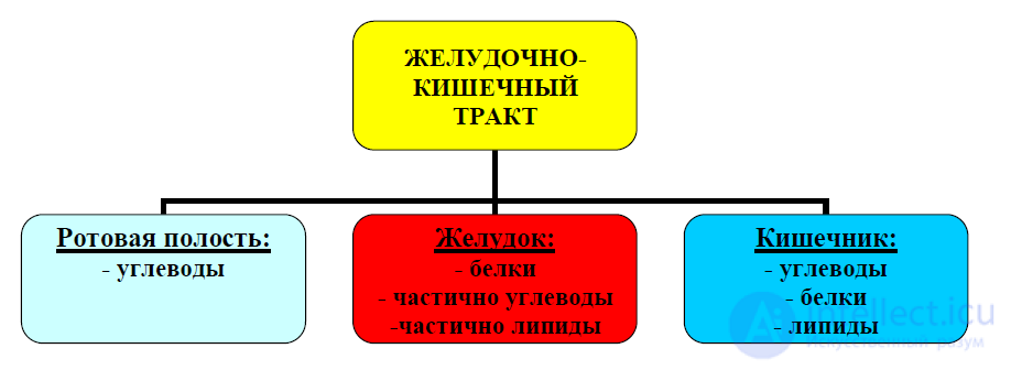
Figure 37 – Digestion of macronutrients in the sections of the GIT
Protein digestion:
gastricsin gastricsin gastricsin chymotrypsin carboxypeptidases
pepsin pepsin pepsin trypsin aminopeptidases
protein → albumoses → peptones → polypeptides → oligopeptides →
dipeptidases
dipeptides → α-amino acids
Carbohydrate digestion (for example, hydrolysis of starch):
amylase amylase maltase
starch → dextrins → maltose → 2 α-glucose
Fat digestion:
lipase lipase
triacylglycerol → diacylglycerol + fatty acid →
lipase
monoacylglycerol + fatty acid → glycerol + fatty acid
1. What is digestion?
2. Which digestion is called cavity (luminal) digestion?
3. Which digestion is called membrane digestion? In which section
of the gastrointestinal tract is it carried out?
4. Name the main cavities of the gastrointestinal tract.
5. List the sections of the small intestine.
6. List the sections of the large intestine
7. What are the enzymes that promote the hydrolysis of proteins called? List them.
8. What are the enzymes that promote the hydrolysis of carbohydrates called? List them.
9. What are the enzymes that promote the hydrolysis of fats called?
10. What is the pH value in the oral cavity?
11. What enzymes are contained in saliva?
12. What digestive processes take place in the oral cavity?
13. Name the pH value in the stomach.
14. What enzymes are contained in gastric juice?
15. What digestive processes take place in the stomach?
16. List the functions of hydrochloric acid in gastric juice.
17. Name the pH value in the intestine.
18. What enzymes are active in the intestine?
19. What digestive processes take place in the small intestine?
20. What is the significance of bile?
21. Name the functions of the large intestine.
22. What functions in the large intestine are performed by the intestinal microflora?

Comments