
Lecture
Spectrophotometry is an analytical technique used across a wide range of industries, providing a quantitative measurement of the interaction of a material under study with electromagnetic radiation. The most commonly used spectrophotometers operate in the ultraviolet and visible light range (hereafter UV/Vis).
Most of the problems associated with the use of spectrophotometry in the visible and ultraviolet regions of the spectrum result from the user's choice of the wrong method or cuvette for the sample. The second problem is the use of an incorrect cleaning strategy. Wherever possible, users should purchase a spectrophotometer optimized for use in a modern research laboratory, so pay attention to the key points for choosing a spectrophotometer listed below.
The spectrophotometric method of detecting the presence and measuring the concentration of substances is based on the fact that most substances have their own specific emission and absorption spectra in one or another region of the spectrum. Therefore, as light passes through a substance, its spectral composition changes. From these changes, the presence of the substance being monitored (the analyte) can be detected (this is qualitative spectral analysis), and by measuring the magnitude of the changes in spectral intensities, the concentration of the analyte or its amount can be calculated (this is quantitative spectral analysis – spectrophotometry).
In practice, spectrophotometric measurements are usually made on plane-parallel transparent solid plates, on thin plane-parallel films containing the analyte, or on transparent solutions poured into plane-parallel cuvettes of calibrated (precisely defined) thickness.
If spectral intensities are measured at only one wavelength, the method is called single-wavelength; if at two wavelengths, dual-wavelength; if at many wavelengths, multi-wavelength. A continuous spectrum of the radiation transmitted through the samples under study may also be recorded. In that case, the entire distribution of spectral intensities within the selected region of the spectrum is used to obtain the analysis results (the continuous-spectrum method).
Depending on the region of the spectrum in which the spectrophotometric measurements are performed, one speaks of infrared, visible, or ultraviolet spectrophotometry.
Let us briefly recall the laws of light absorption and scattering. The basic law of light attenuation as it passes through a substance, in differential form, is:
 (8.1)
(8.1)
where  – the intensity of light in the substance at depth x,
 – the increase in light intensity after passing through a very thin layer of substance of thickness
  ,
,
K– the light attenuation coefficient in the substance. The minus sign means that as light passes through the substance its intensity decreases.
Attenuation of light can occur both due to absorption and due to scattering of light by the substance. The light attenuation coefficient of the substance  generally depends on the wavelength of light λ.
If we integrate the differential expression (8.1) over the thickness of the layer of substance from 0 to d, assuming that at the entrance to the layer (at x=0) the light intensity equals Io, we obtain the integral law of light transmission through a layer of substance:
 (8.2)
(8.2)
The ratio of the intensity of light after passing through the layer of substance to the intensity of the light incident on it
 (8.3)
(8.3)
is called the transmittance of light. Then the integral law of light transmission through a layer of substance is written as:
 (8.4)
(8.4)
The concept of the optical density of a layer of substance, or absorbance, which is the same thing, is also often used.
 (8.5)
(8.5)
Then the integral law of light transmission through a layer of substance is written as:
 (8.6)
(8.6)
When working with plates, thin films, and cuvettes with solution (Fig. 9), reflection of light from their front and back faces must also be taken into account.
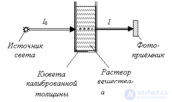
Fig. 9. A typical optical scheme of "transmission" spectrophotometry.
Then the intensity of light passing through the sample under study is described by the well-known formula
 (8.7)
(8.7)
where  – the transmittance coefficient of the cuvette (plate, thin film), accounting only for reflection of light at the faces. Sometimes it also includes the "background" absorption of the pure solvent (the substance of the plate, film) in the absence of the analyte. In optics the analyte is also often called the "dye", since it imparts or changes the color of the solution. In this case the transmittance coefficient
  shows what fraction of the incident light passes through the cuvette with pure solvent (through the plate, film, etc.) in the absence of the analyte.
If we have a solution of only one kind of molecule (one "dye") in a transparent solvent, then

(8.8)
where  – the molar absorption coefficient of the dye; c – its molar concentration.
By measuring the spectral intensities I0 and I in the absorption band of the dye and knowing the transmittance of the cuvette To, and the values  and d, one can calculate the concentration of the dye.
If we have a solution of several different dyes in a solvent that itself partially absorbs light, then

(8.9)
where  – the molar absorption coefficients of the solvent substance and of the 1st, 2nd, ..., n-th dyes respectively;
  – their molar concentrations.
As light passes through a substance it can be attenuated not only by absorption but also by scattering. In that case similar terms due to light scattering appear in formulas (9.2).
Absorption of light energy by a solution. The method is based on the combined Bouguer–Lambert–Beer law, according to which at a given wavelength λ there is a directly proportional relationship between the light absorbance (absorbance Aλ) of a homogeneous solution of a substance, the thickness L of the layer of that solution, and its concentration C. Absorption analysis is carried out using either colorimetric or spectrophotometric instruments.
The fundamental difference between these instruments is that the required photometry wavelength is set by band-pass filters, which transmit a band of wavelengths Δλ, in the first case, and by a monochromator, which emits a flux at a wavelength λ smoothly scanned across the entire range, in the second case..
This type of study is carried out to determine the concentration, size and shape of particles in disperse media.
In general, the phenomenon of light scattering can be described as a change in the characteristics of an optical radiation flux as it interacts with a substance.. Equipment for nephelometric studies consists of specialized photometers for measuring the intensity of light scattered at an angle to the direction of the light flux incident on the solution.
Scattering is caused by optical inhomogeneities in the medium, which may be due either to foreign opaque or semi-transparent particles suspended in the solution, or to density fluctuations of the colloidal-type substance itself, leading to changes in the refractive index.
This type of study of turbid media is based on measuring the change in the intensity of the light energy flux passing through a disperse system. The change in the light energy flux is caused both by absorption and by scattering of the light flux by the disperse system. The method is similar to the colorimetric method.
Luminescence – is the property of a substance to emit energy when its equilibrium state is disturbed by an external excitation source.
Triboluminescence — luminescence that arises upon the fracture of crystalline solids. The causes of triboluminescence vary. In some cases it is explained by the excitation of photoluminescence by electric discharges occurring during the cleaving of a crystalline body, for example after an impact on the substance a faint light is emitted; in other cases it is caused by the movement of dislocations during deformation. For instance, when a sugar crystal is split, a beautiful bluish flash is produced. Luminescence can also occur in ice crystals.
If the excitation is carried out by electromagnetic radiation in the visible or ultraviolet region, this type of luminescence is called photoluminescence.
In turn, photoluminescence is evaluated by the time during which the glow falls to a certain level (usually by a factor of e) after the excitation ceases. Short-lived photoluminescence is called fluorescence. Its duration is determined by the time it takes the molecule to transition from the excited state to the equilibrium state and equals 10-8 – 10-9 sec.. Alongside short-lived, practically instantaneous photoluminescence, there are substances whose emission duration ranges from fractions of a second to several hours. Such photoluminescence is called phosphorescence.
Chemiluminescent analysis methods occupy a special place in luminescence analysis. These are usually oxidation reactions, in the course of which the molecules of the reaction products are excited and light energy is released (radiative deactivation) as they return to the ground state.
Quantitative determination of the content of substances on solid-phase reagent carriers is conventionally called "dry chemistry" systems.
The intensity of the light flux reflected from the colored surface of the carrier is measured, which in turn depends on the concentration of the liquid sample under study.
Flame-photometric and Atomic Absorption Analysis
These types of studies are used mainly for the quantitative determination of metal atoms in solutions from their spectral lines.
At a certain flame temperature, atoms transition from one state to another, which causes the emission or absorption of a light quantum.
Instruments that measure emission are called flame photometers; instruments that measure absorption are called atomic absorption spectrometers.
The polarimetric method of analysis is based on the fact that certain substances, called optically active substances, have the property of affecting polarized light.
As polarized monochromatic light passes through a solution of optically active substances, the plane of polarization is rotated through a certain angle.
The phenomenon of the refraction of light rays at the interface between two optically different media is called refraction (from the Latin "refractus" – refracted).
The refraction of a light ray is determined by the refractive index – the deviation of the ray from its original direction at the interface between two media. This phenomenon is related to the difference in the speed of light propagation in different media.
Exercise 8.1. On what principles is spectrophotometry based? Address the following questions.
Option 1. What is the difference between qualitative and quantitative spectral analysis?
Option 2. What is the difference between single-channel and multichannel spectrophotometric methods?
Option 3. What is the difference between single-wavelength, multi-wavelength and continuous spectrophotometry?
Option 4. What is the light attenuation coefficient? Write the basic law of light attenuation as it passes through a substance in differential form.
Option 5. What is the light transmittance coefficient? Write the law of light transmission through a layer of substance in integral form.
Option 6. How is the attenuation coefficient of light by a solution related to the concentration of each of several different dyes present in it? Write the corresponding formula for the case when the solvent itself also partially absorbs light.
Option 7. What does the law of light transmission through a layer of substance look like in differential form, when the substance does not absorb but only scatters light?
Option 8. What does the law of light transmission through a layer of substance look like in integral form, when the substance does not absorb but only scatters light?
Option 9. What does the law of light transmission through a layer of substance look like in differential form, when the substance both absorbs and scatters light?
Option 10. What does the law of light transmission through a layer of substance look like in integral form, when the substance both absorbs and scatters light?
Exercise 8.2.
Option 1. A cuvette of thickness 2 mm is filled with a dye solution at a concentration of 0.1 mol/l. The molar absorption coefficient of the dye is k = 5 l/(mol·mm). The transmittance coefficient of the cuvette without solution is  . Find the transmittance coefficient of the cuvette with the solution.
Option 2. A cuvette of thickness d mm is filled with a dye solution at a concentration of 0.1 mol/l. The molar absorption coefficient of the dye is k = 5 l/(mol·mm). The transmittance coefficient of the cuvette without solution is  . Find the dependence of the transmittance coefficient of the cuvette with solution on the thickness d.
Option 3. A cuvette of thickness 2 mm is filled with a dye solution at a concentration of 0.1 mol/l. The molar absorption coefficient of the dye is k = 5 l/(mol·mm). The transmittance coefficient of the cuvette with the solution is  , and that of the same cuvette without the solution is
  . Find the concentration of the dye in the solution.
Option 4. A cuvette of thickness 5 mm is filled with a solution of two dyes. One dye is at a concentration of 0.1 mol/l with a molar absorption coefficient k1 = 2 l/(mol·mm), and the second is at a concentration of 0.2 mol/l with a molar absorption coefficient k2 = 0.5 l/(mol·mm). The transmittance coefficient of the cuvette without solution is  . Find the transmittance coefficient of the cuvette with the solution.
Option 5. A cuvette of thickness 5 mm is filled with a solution of two dyes. One dye is at a concentration of 0.1 mol/l with a molar absorption coefficient k1 = 3 l/(mol·mm), and the second is at an unknown concentration with a molar absorption coefficient k2 = 5 l/(mol·mm). The transmittance coefficient of the cuvette without solution is  . Find the concentration of the second dye, if the transmittance coefficient of the cuvette with the solution is
  .
.
Option 6. A cuvette of thickness 5 mm is filled with a dye solution at a concentration of 0.1 mol/l. The molar absorption coefficient of the dye is k = 5 l/(mol·mm). The transmittance coefficient of the cuvette without solution is  . Find the optical density of the cuvette with the solution.
Option 7. A cuvette of thickness 5 mm is filled with a solution of two dyes. One dye is at a concentration of 0.2 mol/l with a molar absorption coefficient k1 = 3 l/(mol·mm), and the second is at a concentration of 0.5 mol/l with a molar absorption coefficient k1 = 0.5 l/(mol·mm). The transmittance coefficient of the cuvette without solution is  . Find the optical density of the cuvette with the solution.
Option 8. A polymer film of thickness 0.2 mm without dye has a transmittance coefficient  . The same film with dye has a transmittance coefficient
  . The molar absorption coefficient of the dye is k = 18 l/(mol·mm). Find the concentration of the dye in the film.
Note
The measuring instruments-sensors reviewed and mentioned within these lectures make up only a small fraction of the overall number of measurement methods and means.

Comments