
Lecture
Any of the molecular properties described above can be used for encoding, transmitting, storing and processing information. But probably the historically first property of molecules that nature put to use billions of years ago, still at the prebiological stage of chemical evolution on Earth, was the property of molecular recognition (eng. – molecular recognition). It was this property that made possible the emergence and subsequent evolution of catalysts – molecules and ensembles of molecules that significantly speed up the course of particular chemical reactions.
The property of molecular recognition is widely used in modern information science as well, first and foremost – in sensorics – the technical science of building and using sensors.
A sensor is a device (unit, instrument, organ) that converts the physical (physico-chemical) properties of an observed object, and their changes, into an information signal for the user.
Sensors are precisely the devices in which information is "born" – information that forms the basis of intelligent behaviour, of the formation and refinement of information models of the surrounding world in "intelligent" systems. After all, any intelligent system, whether it arose naturally or was created artificially, functions successfully and survives in the real world only for as long as it receives objective and high-quality information about that world.
Molecular sensors are very widespread in living nature. But the first artificial sensors of molecular size were created only in the last decades of the 20th century. These are specially synthesized macromolecules that include two important units ( Fig. 3.17). One of them is selectively sensitive to the analyte – molecules of the chemical or biochemical substance whose presence needs to be detected and whose concentration needs to be determined – or to the environmental factors that need to be monitored. This unit is called the recognition element (eng. recognition element). Molecules of natural biochemical enzymes are often used for this purpose. But artificially synthesized recognition elements are also used. The second unit has pronounced luminescent activity, i.e. it glows brightly under the action of short-wavelength external light or another excitation factor. This unit is called the reporter (eng. reporter).
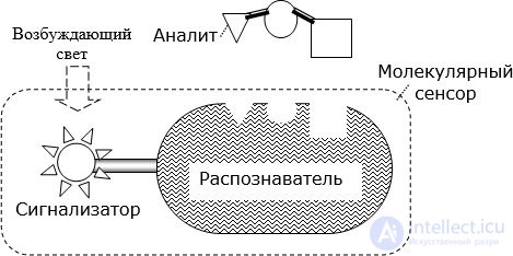
Within the macromolecule these two units interact in such a way that when a particle of the sought analyte attaches to the recognition element, or a specific external factor acts on it, this leads to the quenching or, conversely, the stimulation of the reporter's luminescence.
Thus, the molecular sensor described, by changing the intensity of luminescence, signals the presence and concentration of analyte molecules in the environment, or a change in the characteristics of another specific external factor (for example, temperature, acidity of the medium, etc.). Let us emphasize that each individual macromolecule acts as a full-fledged sensor on its own!
The molecular sensors described are called "sensors with a luminescent marker". While being a full-fledged sensor, they have molecular dimensions and are not designed to work directly with such "users" as a human or a macroscopic instrument. Indeed, the luminescence intensity of a single molecule is relatively weak, which makes it hard to notice against extraneous light interference. Therefore, many (millions, billions) of identical molecular sensors are now used. Their combined luminescent emission makes it possible to build macroscopic sensors.
For example, a luminescent molecular sensor has been synthesized for detecting the presence in the environment of organophosphates, which are found in insecticides and a number of neurotoxins harmful to human health. The unit that selectively recognizes the presence of organophosphates is the enzyme organophosphate hydrolase (eng. organophosphate hydrolase – OPH), and the reporter is carboxynaphthofluorescein (eng. carboxynaphtho-fluorescein – CNF). Using a sensitive photodetector, it was possible to build a portable sensor that reports the presence of organophosphates at concentrations as low as 50 nmol/l. Quantitative measurement of concentration, for example of paraoxon, is performed in the range from 1 to 800 μmol/l.
To effectively exploit the ability of molecules for selective recognition, previously unknown properties of metal nanoparticles (hereafter – NPs) roughly 1–600 nm in size have also been used. As we noted earlier, the electron plasma of such NPs is capable of oscillating at frequencies up to 1016 Hz. Depending on the type of metal and on its own size and shape, an NP has its own characteristic resonant frequencies of electron plasma oscillation. When the frequency of an external electromagnetic field coincides with the resonant frequency, the NPs interact very actively with that field. NPs of metals such as gold and silver, at resonant frequencies, scatter and absorb light much more strongly than molecules of even the best organic dyes. Thanks to this, the places where gold or silver nanoparticles accumulate become intensely coloured and immediately noticeable.
Another valuable property of gold and silver NPs is that they allow targeted modification of their outer surface. In order to build sensors that use the property of molecular recognition, the NP surface is first chemically "passivated" by depositing a thin layer of hydrogel onto it. These are short (up to 10-20 carbon atoms in the chain) linear polymer molecules (thiols, disulfides, thioethers) that readily form strong covalent bonds with gold and silver. In solution, such molecules quickly attach to the surface of the nanoparticles, forming a self-assembled monolayer (eng. selfassembled monolayers – SAM) with fairly dense packing ( Fig. 3.18). With water molecules this layer forms a hydrogel, which reliably protects the metal surface from interaction with other molecules. Afterwards, recognition-element molecules are chemically deposited onto this thin hydrogel film.
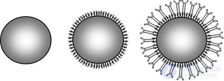
Metal NPs prepared in this way are called "selective", and if the recognition-element molecules are of biological origin, then "bioselective".
Bioselective NPs have been used for several decades already in so-called immunochromatographic analysis. Fig. 3.19 shows the construction of an immunochromatographic test strip used in such an analysis.
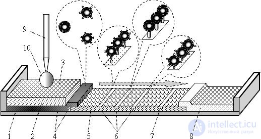
On the plastic base 1 there is a filter 2 with a receiving zone 3, beneath which is a pad soaked with a gel 4 containing bioselective NPs. Here there may be NPs of one kind, designed to recognize a single analyte, or bioselective NPs of two or three kinds (each designed for its own analyte). Pad 4 is in contact with chromatographic membrane 5, most often made of nitrocellulose. Along this membrane a drop of liquid can seep through, spreading as in a chromatography column. In certain places the membrane is crossed by transverse strips 6, onto which antibodies to the corresponding analyte are immobilized. These places are called test zones or binding zones. The number of test zones (1-3) equals the number of analytes being monitored simultaneously. In control zone 7, antibodies to all the bioselective nanoparticles are immobilized, especially to those to which no analyte has attached. And at the end of membrane 5 is the absorption zone 8.
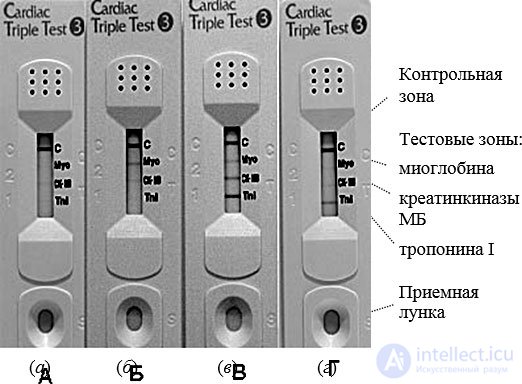
The operation of the test strip will be explained using the example of the three-component "ImmunTech" cardio-test made by YD Diagnostics, South Korea. Fig. 3.20 shows their appearance. On the plastic cover, next to the control zone and the test zones, corresponding abbreviated labels are printed, deciphered in the figure on the right. Above the receiving zone a well is formed, next to which there is an embossed label "S" (eng. Sample). Above the absorption zone there are many holes that allow the liquid to evaporate freely.
The sensor uses bioselective gold NPs of three kinds. Immobilized on the surface of these NPs are molecules that selectively recognize:
troponin I (we will call these bioselective NPs of the 1st kind);
creatine kinase MB (we will call these bioselective NPs of the 2nd kind);
myoglobin (we will call these bioselective NPs of the 3rd kind).
The molecules of all three of these substances are called "cardiac markers", since their appearance in the patient's blood indicates specific changes in the tissues of his heart.
In the test zones, antibodies are immobilized that have a strong chemical affinity for one of the above-mentioned substances (antibodies of the 1st, 2nd and 3rd kind respectively). When a couple of drops of the patient's blood are placed in the receiving well, the liquid portion (plasma) of this blood seeps through filter 2 (here and further in this paragraph see Fig. 3.19) and enters pad 4. Here the blood plasma begins to interact with the bioselective gold NPs. If myoglobin molecules are present in the blood plasma, they attach to the bioselective NPs of the 1st kind. If creatine kinase MB molecules are present, they attach to the bioselective NPs of the 2nd kind. And if troponin I molecules are present, they attach to the bioselective NPs of the 3rd kind. The footnote on the left conditionally shows NPs of the three kinds with analyte molecules attached to them. Next, under the action of capillary forces, the mobile liquid phase begins to seep and move along chromatographic membrane 3. The direction of movement is shown in Fig. 3.19 by a grey arrow. When the mobile liquid phase reaches the first test zone 6, the bioselective NPs of the 1st kind are "captured" by the 1st-kind antibodies immobilized there. And if there are many troponin I molecules in the blood sample, the first test zone turns red, the colour characteristic of gold nanoparticles. Meanwhile the mobile liquid phase continues to move further. When it reaches the second test zone, the 2nd-kind antibodies immobilized there capture the bioselective NPs of the 2nd kind, to which creatine kinase MB has attached. And if there is a large amount of creatine kinase in the blood sample, the second test zone also turns red. When the mobile liquid phase reaches the third test zone, the 3rd-kind antibodies immobilized there capture the bioselective NPs of the 3rd kind, to which a myoglobin molecule has attached. And if there are many myoglobin molecules in the blood sample, the third test zone also turns red. The mobile liquid phase then reaches the control zone 7, where the antibodies immobilized there capture all the gold NPs not captured earlier. And the control zone turns red. The mobile liquid phase then reaches the absorption zone 8, from which it can evaporate freely.
Thus, the control zone is coloured after chromatography almost always, while the test zones are coloured depending on the presence of the corresponding cardiac markers in the blood sample.
Fig. 3.20 shows 4 typical cases. In case (a), intense colouring is observed only in the control zone C. Since none of the cardiac markers has been detected, there is neither infarction nor a risk of infarction. In case (b), the presence of myoglobin and a small amount of creatine kinase MB has been detected in the blood sample. This indicates to the cardiologist that the patient's risk of myocardial infarction is quite high. In case (c), the characteristic picture of myocardial infarction is observed: all three cardiac markers are present, with an especially large amount of troponin I. In case (d), the troponin test zone is coloured and the creatinine zone is much weaker. For the cardiologist, this is evidence that a myocardial infarction has occurred, and that it occurred more than 4 days ago.
Thus, the control zone is coloured after chromatography almost always, while the test zones are coloured depending on the presence of the corresponding cardiac markers in the blood sample.
The use of such immunochromatographic tests by ambulance crews, in clinics, and by family doctors makes it possible to reduce the number of diagnostic errors many times over, to save many lives, and to provide patients with qualified medical care in a timely manner.
Immunochromatographic tests have already been developed and are in use for detecting more than 150 types of diseases, for assessing the presence in human blood of a wide range of hormones, antibodies, enzymes, and narcotic substances. Since only blood is needed for the analysis, the analysis can be performed anonymously. It can be carried out at home or even while travelling.
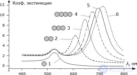
Another practically important property of gold and silver NPs is that their absorption and scattering spectrum depends significantly on the aggregation of NPs into clusters. Fig. 3.21 shows, for example, the changes in the extinction spectrum (a measure of the rate of light attenuation) of an emulsion with silver nanoparticles 20 nm in diameter as the NPs combine into aggregates. Since an additional, very strong light-absorption band appears upon aggregation (coagulation) of the NPs, the colour of the solution containing the NPs changes substantially.
Sensors that use molecular recognition and the change in colour of NPs are called colorimetric nanosensors.
Let us consider one example of such sensors, designed to detect the presence in solution of specific oligomers – molecules made up of a relatively small number of amino acids. For this purpose, molecules of 5'- or 3'-mercaptoalkyl-oligonucleotide, containing 28 amino acids (amino-acid residues), are immobilized on gold NPs 13±2 nm in diameter after passivation of their surface with thiol. Of these, the last 15 residues are intended for the selective recognition of the given oligomers. They are exactly complementary to the oligomers whose presence needs to be detected. These are the recognition units. Since they are not complementary to one another, they prevent coagulation of the NPs. Therefore the initial solution has a pronounced light-absorption band near a wavelength of 520 nm and appears red.
When molecules of a complementary oligomer appear in the solution, they attach to the recognition units on the NPs, as a result of which coagulation of the NPs becomes possible. This process is shown schematically at the top of Fig. 3.22. Coagulation leads to substantial changes in the transmission spectrum of the solution, shown at the bottom of Fig. 3.22. After being held for several hours, the colour of the solution changes from red to red-violet. This significant change in colour makes it possible to see the presence of the analyte in the solution "with the naked eye", without any instruments.
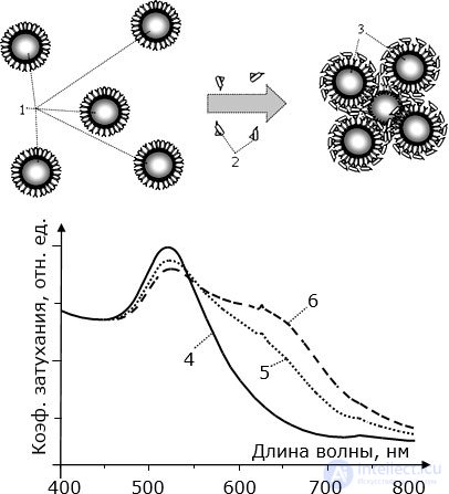
Control experiments show that the presence in solution of molecules of other oligomers, even ones very similar to the analyte but not complementary to the recognition units, does not lead to coagulation of the NPs or to a change in the colour of the solution.
The detection threshold for the complementary analyte is on the order of 10 fM (10-14 mol/l).
In the same way as described above, molecules oriented toward the recognition of telomerase can be attached to gold NPs. This specific enzyme, discovered in 1984, extends repeating DNA fragments ("TTAGGG") onto the telomeres located at the 3'-end of the DNA of eukaryotic cell chromosomes. It is precisely this that, as it turns out, makes vitally important cells (germ cells and stem cells) "immortal", allowing them to divide for a very long time. Telomerase is also present in the cancer cells of most types of cancer (up to 85%). Therefore, by detecting the presence of telomerase in living tissues in which germ cells or stem cells are known to be absent, one can also diagnose the presence of cancer cells.
It has been shown experimentally that, using specialized gold NPs and the colorimetric method, one can experimentally detect with the naked eye the presence of telomerase secreted by just a dozen cancer cells. And, using spectrophotometric devices, one can detect telomerase molecules secreted even by a single cancer cell. The time required for detection is on the order of 10 minutes.
Sensory "nanoprobes" similar to those described (gold NP – thiol – recognition-element molecule) are already widely used for detecting the presence of dozens of important biological molecules in vivo at fairly low concentrations, using minimal sample volumes.

Comments