
Lecture
Vitamin-like substances, also known as growth factors, cofactors, or biologically active compounds, are chemical compounds that have functions similar to those of vitamins but are not true vitamins, because the body can synthesize them on its own or obtain them from food in sufficient amounts.
Vitamin-like substances are a group of organic compounds that have high biological activity and are similar to vitamins but, unlike true vitamins, most of them can be synthesized in the human body in the required amounts during normal metabolism. Although many of them have been called vitamins, their classification as vitamins is disputed. This group includes: rutin (vitamin P), inositol (vitamin B8), carnitine (vitamin Bm, B11), xanthopterin (vitamin B14), lipoic acid (vitamin N), orotic acid (vitamin B13), pangamic acid (vitamin Q), methylmethionine sulfonium chloride (vitamin U), and choline (B4 or Bp).
These substances are important for maintaining health and normal body function, but their presence in food or supplemental sources is usually not considered essential for life, as is the case with vitamins and minerals. However, some people may benefit from taking these substances as dietary supplements, especially if they have a deficiency or an increased need for them. It is important to consult a physician or a nutrition specialist before starting any supplements.
| Letter designation (obsolete ones in parentheses) | Chemical name according to international nomenclature (other names in parentheses) | Solubility (F — fat-soluble W — water-soluble) |
Consequences of deficiency (avitaminosis), physiological role | Tolerable upper intake level | Daily requirement |
|---|---|---|---|---|---|
| These substances were formerly considered vitamins or were vitamin candidates, but are no longer classified as such. | |||||
| (B4) | Choline | W | Precursor of the neurotransmitter acetylcholine. Deficiency leads to fat deposition in the liver, renal failure, and bleeding. | 20 g | 425—550 mg |
| (B8) | Inositol (inosite, myo-inositol) | W | No data | No data | No data |
| (B10) | 4-Aminobenzoic acid (p-aminobenzoic acid, para-aminobenzoic acid, PABA) | W | Stimulates the production of vitamins by the intestinal microflora. | No data | Not established |
| (B11, BT) | Levocarnitine | W | Disturbances of metabolic processes | No data | 300 mg |
| (B13) | Orotic acid | W | Various skin diseases (eczema, neurodermatitis, psoriasis, ichthyosis) | No data | 0.5—1.5 mg |
| (B15) | Pangamic acid | W | No data | No data | 50—150 mg |
| (N) | Lipoic acid, thioctic acid | F | Necessary for normal liver function | 75 mg | 30 mg |
| (P) | Bioflavonoids, polyphenols | W | Capillary fragility | No data | No data |
| (U) | Methionine S-methylmethionine sulfonium chloride | W | Anti-ulcer factor; vitamin U (from Latin ulcus — ulcer) | No data | No data |
Vitamin P (from permeability) is a large group of polyphenolic compounds, united under the common name "bioflavonoids", that have a vessel-strengthening effect similar to that of vitamin C. Because of their close association with vitamin C, bioflavonoids are sometimes called vitamin C2. Other names for vitamin P are rutin and thioctic acid.
Chemically, bioflavonoids are derivatives of chromone or flavone. They contain a diphenylpropane skeleton (C6–C3–C6), consisting of two aromatic rings connected by a three-carbon fragment that forms a pyran or pyrone ring (when a double bond is present), and they have the general formula
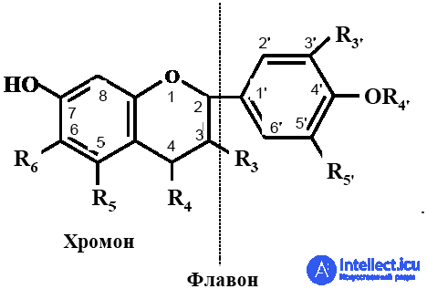
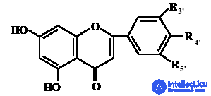
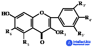
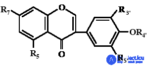
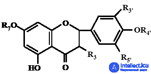
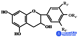
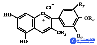
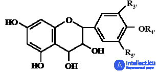
They differ from one another in the degree of oxidation of the pyran ring, as well as in the number and position of the hydroxyl groups in the aromatic rings. The flavonoids most commonly found in nature have four (positions C5, C7, C3', C4') or five (positions C5, C7, C3', C4', C5') hydroxyl groups in the molecule; the maximum number of hydroxyls is seven. In structure, flavonoids resemble tocopherols in which the isoprenoid chain is replaced by a phenolic radical containing hydroxyl groups. The antioxidant properties of flavonoids are associated with the presence of hydroxyl groups at positions C3'–C5', and the acidic properties with the hydroxyl at C7. Depending on the nature of the substituents in the ring, seven main groups of flavonoids are distinguished (Table 2.3.1).
Table 2.3.1. Main groups of flavonoids with P-vitamin activity and their aglycones
|
Structural formula of flavonoids |
Name of the flavonoid group and corresponding aglycones |
|
|
Flavones: apigenin |
|
|
Flavonols: |
|
|
Isoflavones: |
|
|
Flavanones: Flavanonols: |
|
|
Flavanols (catechins): |
|
|
Anthocyanidins: |
|
|
Leucoanthocyanidins: |
The different groups of flavonoids are not equally widespread in nature: about 40% of them are flavonols, flavones are less common, and flavanones are even rarer. In nature these groups of flavonoids are interconvertible, so in the plant kingdom they occur together (Table 2.3.2). Many of these compounds, which have a broad spectrum of action (antiseptic and antioxidant properties), possess P-vitamin activity to varying degrees.
Table 2.3.2. Content of compounds with P-vitamin activity in fruits and vegetables
|
Name of |
Name of |
Vitamin P content |
|
|
Fresh tissue weight, mg % |
Dry tissue weight, % |
||
|
Apples |
Catechins |
100–150 |
|
|
Leucoanthocyanins |
450–600 |
||
|
Pears |
Leucoanthocyanins |
100–250 |
|
|
Quince |
Leucoanthocyanins |
200–825 |
|
|
Sour cherry |
Anthocyanins |
1300–2500 |
|
|
Dark sweet cherry |
Anthocyanins |
225–900 |
|
|
Plum |
Leucoanthocyanins |
0.9 |
|
|
Oranges |
Hesperidin |
3.07 |
|
|
Lemons (juice) |
Eriodictyol |
20 |
|
|
Quercetin |
22 |
||
|
Hesperidin |
1.4 |
||
|
Pomegranates |
Anthocyanins |
200–700 |
|
|
Grapefruit: |
|||
|
whole fruit |
Naringin |
120–360 |
|
|
Juice |
Naringin |
36.0 |
|
|
Naringenin |
12.4 |
||
|
Neohesperidin |
1.1 |
||
|
Hesperidin |
1.0 |
||
|
Black |
Rutin |
3.5 |
|
|
chokeberry |
Quercetin |
1.8 |
|
|
Hesperidin |
0.9 |
||
|
Anthocyanins |
4000 |
||
|
Sea buckthorn |
Rutin |
2.3 |
|
|
(fruits) |
Hesperidin |
1.3 |
|
|
Rose hips |
Quercetin |
2.8 |
|
|
(fruits) |
Hesperidin |
1.8 |
|
|
Black currant |
Anthocyanins |
1000–1500 |
|
|
Onion: |
|||
|
Bulb |
Rutin |
0.95 |
|
|
Skin |
Rutin |
2.4 |
|
|
Quercetin |
1.3 |
||
|
Red pepper |
Rutin |
2.1 |
|
|
(powder) |
Quercetin |
0.95 |
|
|
Hesperidin |
1.2 |
||
|
Sorrel |
Rutin |
500 |
|
|
Dill |
Quercetin |
170 |
|
|
Parsley (leaves) |
Apigenin |
157 |
Flavonoids are plant pigments that determine the color of various plant organs, in particular flowers and fruits. They are found much less often in lower plants, insects, and microorganisms. Young flowers and unripe fruits are the richest in flavonoids, which are localized in the cell sap of plants in dissolved form. The distribution of flavonoids in the pulp and skin of fruits and berries varies: in some it is relatively uniform, while others contain them only in the skin, for example, apples.
Most bioflavonoids occur in plants bound to sugars (flavonoid glycosides) and organic acids, and less often in the free state (flavonoid aglycones). For example, the flavonoid glycosides hesperidin and naringin contain hesperetin and naringenin, respectively, as their aglycones; the aglycone of rutin is quercetin, and anthocyanidins are the aglycones of anthocyanins. Because of their low solubility, flavonoid aglycones are located in the lipid parts of plants — fat droplets and wax layers.
The main dietary sources of flavonoids are fruits, vegetables, and beverages (tea, juices, wine). For example, the flavonoid content of red wine is higher than that of grape juice, owing to the microorganisms involved in winemaking. In foods, flavonoids can be present in monomeric, dimeric, and polymeric forms; the latter are called tannins.
The biological role of flavonoids consists in stabilizing the intercellular matrix of connective tissue and reducing capillary permeability. The strength of the walls of blood capillaries is directly controlled by the hormones of the adrenal cortex; the role of P-vitamin substances is that they protect the adrenal medullary hormone adrenaline (epinephrine) from oxidation, prolonging its action.
P-vitamin activity is manifested through effects on certain enzyme systems of the body that regulate vascular permeability. For example, bioflavonoids, together with vitamin C, inactivate the enzyme hyaluronidase, which catalyzes the breakdown of the heteropolysaccharide hyaluronic acid, the main substance of connective tissue. An inhibitory effect of vitamin P on ascorbate oxidase has also been shown. The vitamin protects ascorbic acid from oxidation and promotes the regeneration of dehydroascorbic acid with the participation of glutathione and the absorption of vitamin C in the human body.
The antioxidant action of flavonoids is associated with their ability to block the catalytic action of heavy metals by binding them into stable complexes. Flavonoids are thought to act like other lipophilic antioxidants, mimicking their action to some extent.
Bioflavonoids are not synthesized in the human body and must be supplied with food. A deficiency of vitamin P can lead to hemorrhages in the mucous membranes and skin and to bleeding gums because of the fragility and increased permeability of blood vessels. Vitamin P deficiency leads to rapid fatigue. The exact daily requirement for vitamin P in humans is unknown; 25–50 mg/day is taken as the physiological norm. No cases of hypervitaminosis P are known to date; flavonoids are nontoxic, and any excess is easily eliminated from the body.
Examples of the most thoroughly studied and significant bioflavonoids for humans are tea catechins, rutin, quercetin, and dihydroquercetin, which are produced on an industrial scale; hesperidin and naringin are also manufactured. The chemical synthesis of some bioflavonoids and their analogues (rutin, esculetin) has been achieved.
Rutin, or quercetin 3-rutinoside (3-rhamnoglycosyl-3,5,7,3',4'-pentahydroxyflavone), is a flavone derivative (a flavonol). Hydrolysis of the rutin molecule and cleavage of the sugar residue yields the aglycone quercetin, which has P-vitamin activity.
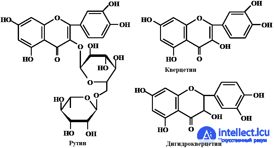
Together with vitamin C, rutin takes part in oxidation-reduction reactions, stimulates tissue respiration, regulates the permeability of capillary vessels, increasing their strength and preventing sclerotic damage, and protects ascorbic acid from oxidation. Rutin and quercetin are powerful antioxidants: they can not only bind free oxygen-containing radicals but also inhibit their formation by binding transition metals. For example, the complex of rutin with iron (II) ions is almost five times more active than rutin itself and acts as an effective trap for the superoxide radical, whose formation initiates the process of lipid peroxidation of biomembranes.
Rutin is usually found in the same plant foods that contain vitamin C. The most rutin is found in black currants, rose hips, citrus fruits, green tea, lettuce, buckwheat flowers and leaves, grapes, cabbage, apples, and plums. Rutin is obtained from buckwheat green mass and from the buds of the Japanese pagoda tree (Sophora japonica).
Dihydroquercetin. Another promising natural antioxidant is dihydroquercetin (DHQ, taxifolin, 3,3',4',5,7-pentahydroxyflavanone), which belongs to the group of bioflavonoids and is extracted on an industrial scale from larch wood. In antioxidant action DHQ is comparable to α-tocopherol but surpasses it in stability. The range of applications of DHQ is quite broad; it is used in the food, cosmetic, and pharmaceutical industries. In the food industry, DHQ and quercetin are used as harmless antioxidant additives to extend the shelf life (by 2–2.5 times) of lipid-containing foods (vegetable oils, pork and milk fat, powdered milk, palm oil).
METHODS OF DETERMINING FLAVONOIDS
|
Parameter |
Characteristic |
|
|
Rutin |
Quercetin |
|
|
Formula |
C27H30O16×3H2O |
C15H10O7·3H2O |
|
Molecular weight |
610 |
302 |
|
Melting point, °C |
182–192 |
316–317 |
|
Solubility |
Freely soluble in methanol and isopropanol, partially soluble in acetone |
Soluble in acetone, ethanol, methanol, and isopropanol |
|
Absorption maximum, λmax, nm |
257, 362 |
256, 375 |
|
Specific absorbance coefficient, ε1cm1%, ml/µg×cm |
32.5 |
In pure form, flavonoids are crystalline compounds that are yellow (flavones, flavonols, etc.), colorless (isoflavones, catechins, flavanones, flavanonols), or colored red or blue (anthocyanins) depending on the pH of the medium. In an acidic medium anthocyanins are shades of red or pink; in an alkaline medium, blue.
Flavonoid glycosides containing more than three sugar residues are soluble in water but insoluble in diethyl ether and chloroform. Acid or enzymatic hydrolysis is characteristic of flavonoid glycosides. Flavonol 3-glycosides are readily hydrolyzed on heating in 0.1–1% solutions of mineral acids, whereas flavone 7-glycosides are hydrolyzed only on heating for several hours in 5–10% acid solutions. Flavonoid C-glycosides are hydrolyzed only by a mixture of concentrated hydrochloric and acetic acids. Flavonoid aglycones dissolve in diethyl ether, acetone, and alcohols and are practically insoluble in water.
Flavonoids are usually extracted with alcohols (ethyl, methyl). The alcohol extract is evaporated, and the residue is dissolved in hot water. Nonpolar compounds are washed out of the aqueous phase with chloroform or carbon tetrachloride, and the flavonoids are extracted successively with diethyl ether (aglycones), ethyl acetate, and butanol (glycosides).
Chromatography is used for further separation of the flavonoid fractions. They are readily separated by chromatography and are characterized by differences in color in visible and ultraviolet light. To visualize flavonoids after separation, the most commonly used agents are ammonia vapor, solutions of alkali in methanol, aqueous solutions of aluminum chloride, a solution of antimony pentachloride in carbon tetrachloride, and a solution of potassium or sodium borohydride in isopropyl alcohol, followed by spraying with an alcoholic solution of aluminum chloride and heating at 120 °C. Flavonoids are identified using UV, IR, and NMR spectroscopy.
The most common methods of quantitative analysis of bioflavonoids are chromatographic and spectrophotometric methods and densitometry combined with chromatography. Photocolorimetric methods of determining flavonoids are based on color reactions with metal salts (aluminum, zirconium, titanium, chromium, antimony) and with citric-boric reagent.
Choline (vitamin B4 or Bp) is 2-hydroxyethyl(trimethyl)ammonium hydroxide; its name comes from the Greek word chole, meaning bile.
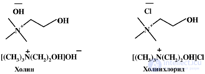
It is freely soluble in water, is a strong base, and forms salts with acids; the most common salt is choline chloride, which is widely used in pharmacology. Choline is a component of the most important phospholipids — lecithins and sphingomyelins — which are lipid components of cell membranes and make up half of the sensitive myelin sheath in nerve cells.
Not all mammals are able to synthesize vitamin B4, but humans, some animals, and microorganisms are capable of producing choline. In mammals, choline synthesis proceeds stepwise in the liver from phosphatidylserine, with methionine acting as the donor of methyl groups. An additional amount of choline is synthesized by the human intestinal microflora with the participation of the amino acids methionine and serine and of vitamins B12 and B9, reducing the share of endogenous choline to one fifth of the daily requirement for it. Strictly speaking, choline is not a vitamin, since it is synthesized in the body and used as a structural substance in building cell membranes. However, because the synthesis of methyl groups is limited, choline is classed as a pseudovitamin, and the bulk of it must be supplied with food.
Rich sources of choline are animal and plant tissues; some microorganisms also produce choline. Important dietary sources include eggs, liver, soy, all kinds of cabbage, peanuts, meat, and fish (Table 2.3.4).
Table 2.3.4. Choline content of foods
|
Name of product |
Mass fraction, mg/g |
|
Chicken egg yolk |
1713 |
|
Pork liver |
552 |
|
Chicken egg |
504 |
|
Sprouted wheat |
406 |
|
Beef kidneys |
333 |
|
Sprouted rice |
300 |
|
Soy (beans) |
300 |
|
Pork kidneys |
256 |
|
Cabbage |
251 |
|
Spinach |
238 |
|
Lentils |
223 |
|
Cod |
200 |
|
Salmon |
181 |
|
Oatmeal |
156 |
|
Pearl barley |
139 |
|
Herring |
127 |
|
Tomatoes |
106 |
|
Meat |
75–100 |
|
Potatoes |
66 |
|
Beef kidneys |
63 |
|
White bread |
62.5 |
|
Black bread |
56.5 |
|
Cabbage |
23 |
|
Spinach |
22 |
|
Milk |
14.7 |
|
Orange juice |
12 |
Plant foods contain less choline than foods of animal origin, but green leaves and legumes are good sources among them. Choline is found in the germ of cereals, in hawthorn fruits, cabbage, spinach, and soy. Losses of choline when cooking plant foods range from 10–40%, when cooking meat and liver they are 18%, and when baking the losses are insignificant.
The main metabolic form of choline is lecithin, which promotes lipid metabolism in the liver, that is, it has hepatoprotective properties. Choline prevents fatty liver, because when vitamin B4 is lacking, the release of fatty acids from the liver slows down. In addition, the body synthesizes acetylcholine from choline — the most important neurotransmitter, which takes part in the transmission of nerve impulses; hence the role of choline in improving memory. Choline is a methylating agent and takes part in biochemical methylation reactions, namely in the synthesis of the essential amino acid methionine.

The methylation of homocysteine by choline is a reversible reaction, and when choline is lacking in the body, it is formed from the essential amino acid methionine. Nevertheless, biological synthesis cannot fully meet the body's need for choline. Given that the formation of endogenous choline depends on the supply of methionine and vitamins (B9, B12), it is evident that choline deficiency is possible in humans as a secondary deficiency associated primarily with protein deficiency. Choline deficiency combined with protein deficiency can cause fatty degeneration of the liver and cirrhosis. The human requirement for choline is 1.5–4 g/day, and up to 6 g/day under severe stress.
Choline, its salts and esters are used in the food industry as an emulsifier and are registered as the food additive E1001; as a component of lecithin, as an antioxidant — E322.
1. Inositol (B8, myo-inositol, meso-inositol) is cyclohexane-1,2,3,4,5,6-hexol — cis-1,2,3,5-trans-4,6-cyclohexanol — a biologically active compound. The melting point of inositol is 225 °C. Its crystals are readily soluble in water (10–14 g/100 mL). Inositol is resistant to acids and alkalis.
In terms of its spatial structure, inositol can exist as eight cis-/trans-stereoisomers, seven of which are optically inactive (meso forms), while one exists as two optical isomers (D and L) and one racemate. Only one of these isomers has vitamin activity and it is called myo-inositol.
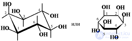
2. The cyclohexane ring of myo-inositol has a chair conformation in which the hydroxyl groups at C1–C3 and C5 lie on one side of the ring and are designated cis-hydroxyls, while the remaining ones lie on the other side and are designated trans-hydroxyls. The metabolically active form of myo-inositol is phosphatidylinositol. Vitamin activity is exhibited by the salts of inositol phosphoric acid — phytins — which have the same biological effect as myo-inositol. The natural inositols include only myo-inositol, D-inositol and L-inositol.
Inositol is a universal component of most living organisms and occurs both in the free and the bound state, in the form of phosphoric esters, phospholipids and phosphoproteins. Inositol is synthesized in mammals from glucose in the liver, by cyclization of glucose-6-phosphate to inositol-1-phosphate with subsequent dephosphorylation in many tissues and organs (heart, liver, kidneys).
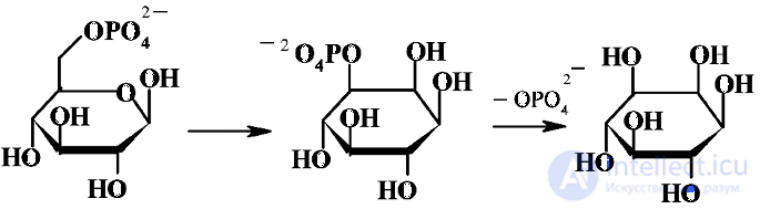
With the bloodstream it reaches all cells that cannot produce it themselves. An additional source of inositol is its synthesis in the intestine. Inositol is found in high concentrations in brain cells, where it accumulates in biomembranes; it is necessary for the functioning and growth of nerve cells. There is a great deal of inositol in the lens, the posterior wall of the eye and the tear fluid, so a deficiency of this vitamin can lead to various eye diseases.
3. In foods, inositol is present in three forms: inositol, phytic acid and inositol-containing phospholipids. Rich sources of inositol are plant seeds, malt, and also citrus fruits, cabbage, carrots, beets, potatoes, tomatoes and strawberries, in which it is present mainly as phytic acid, which can convert calcium, magnesium, iron and zinc into unabsorbable salts, complicating their absorption (Table 2.3.5). In addition, the assimilation of phytin from plant foods requires a large amount of the enzyme phytase, which is contained in gastric juice and produced in the intestine, so impaired digestion can lead to inositol deficiency. In foods of animal origin, inositol is found predominantly in the form of phospholipids.
4. The biological role of inositol is associated with phospholipid metabolism and the formation of inositol-1,4,5-trisphosphate, an active second messenger of intracellular signals. Inositol has a lipotropic effect, lowers blood cholesterol levels, prevents hardening of the arteries, restores the structure of nervous tissue, normalizes sleep, and stimulates the motor function of the digestive tract. In medical practice, inositol is used in liver diseases, and phytin is used as a source of phosphorus in nervous diseases and to stimulate hematopoiesis.
Table 2.3.5. Inositol content in foods, mg/g
|
Food product |
Mass fraction, mg% |
|
Asparagus |
0,29–0,68 |
|
White beans |
2,83–4,40 |
|
Broccoli |
0,11–0,30 |
|
Cabbage |
0,18–0,70 |
|
Carrots |
0,52 |
|
Cauliflower |
0,15–0,18 |
|
Trout |
0,11 |
|
Peas |
1,16–2,35 |
|
Spinach |
0,06–0,25 |
|
Tomatoes |
0,34–0,41 |
|
Milk |
0,04 |
|
Egg |
0,09 |
|
Egg yolk |
0,34 |
|
Apples |
0,10–0,24 |
|
Grapes |
0,07–0,16 |
|
Grapefruit |
1,17–1,99 |
|
Oranges |
3,07 |
|
Pear |
0,46–0,73 |
|
Rice |
0,15–0,30 |
|
Wheat |
1,42–11,5 |
|
Beef |
0,09–0,37 |
|
Chicken meat |
0,30–0,39 |
|
Pork |
0,14–0,42 |
|
Beef liver |
0,64 |
|
Chicken liver |
1,31 |
|
Tuna |
0,11–0,15 |
The daily requirement for inositol is 1–1.5 g/day; about ¾ of the daily requirement is produced by the body from glucose, and the remainder comes from food. In most cases, poor nutrition leads to disturbance of the intestinal microflora and thereby to inositol deficiency.
Ubiquinones (coenzyme Q, CoQn). The name comes from the Latin word ubique — everywhere, anywhere — and quinone. In 1978 the American scientist P. Mitchell received the Nobel Prize for developing the theory of the action of coenzyme Q10, an indispensable cellular component that takes part in ATP synthesis. Structurally, they are derivatives of 2,3-dimethoxy-5-methyl-1,4-benzoquinone with an isoprenoid chain at position C6:
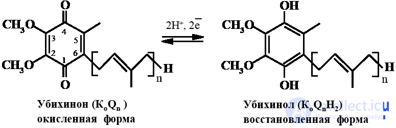
They differ from one another in the number of isoprene units, from 6 to 15. Natural ubiquinones most often contain from six to ten isoprene units (Q6–Q10). At n=1–6 ubiquinones are oily red substances; at n=7–15 they are yellow or yellow-orange crystals that are insoluble in water but soluble in organic solvents. In nature, ubiquinones occur in all organisms — mammals, fungi, bacteria, protozoa and insects. They are present in small amounts in plant mitochondria. Ubiquinones isolated from bacteria contain from six to nine isoprene units, those of higher plants nine, and those of mammals nine to ten. Coenzyme Q10 is present in the human body.
Ubiquinones are capable of reversible redox transformations and are localized in the inner mitochondrial membranes. Their main function is coenzymatic and is associated with the transfer of electrons and protons during respiration and oxidative phosphorylation. Since coenzyme Q10 takes part in supplying cells with energy, in humans the largest amount of coenzyme Q10 is concentrated in the mitochondria of cells of the most energy-consuming organs — the heart, liver and pancreas.
Another important function of ubiquinones is antioxidant. Together with α-tocopherol, β-carotene and selenium, they are powerful antioxidants and are consumed first. Unlike the antioxidants named above, ubiquinones are synthesized in the human body itself and are able to continuously restore their antioxidant activity, passing from the oxidized form (ubiquinone) to the reduced form (ubiquinol). According to some data, they play an important role in protecting low-density lipoproteins from oxidation. In heart cells, Q10 reduces the level of free radicals and slows the development of atherosclerosis.
In animal cells, ubiquinones are synthesized in sufficient amounts from the corresponding precursors. Ubiquinone synthesis is believed to begin in the endoplasmic reticulum and end in the Golgi complex, from which they are distributed to all organelles. Formation of the isoprenoid chain shares common steps with cholesterol synthesis, while the benzene portion is formed from tyrosine. Therefore, ubiquinone synthesis may be limited by the availability of tyrosine and its precursor, the essential amino acid phenylalanine.
With age, the ubiquinone content of human tissues declines. A 75% ubiquinone deficiency causes cell death, and a 25% reduction in level noticeably affects the function not only of internal organs (heart, kidneys) but also of the nervous and endocrine systems. Coenzyme Q10 deficiency leads to diseases such as heart failure, loss of immunity, muscular dystrophy and chronic liver damage.
Although only 2–3% of exogenous Q10 is absorbed in the stomach, it is widely used in the treatment of atherosclerosis. Data on dietary sources of ubiquinones in the literature are inconsistent (Table 2.3.6). However, since ubiquinones are present in all mitochondrial membranes, it may be assumed that many foods are rich in them, especially heart and muscle tissue.
Table 2.3.6. Coenzyme Q10 content in foods
|
Food product |
Mass fraction, mg% |
|
Pork heart |
20,30 |
|
Beef heart |
4,10 |
|
Herring |
2,70 |
|
Beef liver |
1,90 |
|
Chicken legs |
1,70 |
|
Trout |
1,10 |
|
Leg of lamb |
0,29 |
|
Spinach |
0,23 |
|
Oranges |
0,22 |
|
Cauliflower |
0,06 |
Lipoic acid (thioctic acid, vitamin N) is structurally a thio-derivative of valeric acid and readily undergoes redox transformations:
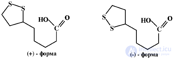
Lipoic acid exists as two stereoisomers, of which the dextrorotatory one is biologically active. The natural, dextrorotatory form of lipoic acid melts at 47.5 °C, [a] +104, while the synthetically obtained form is the D,L-racemate with a melting point of 60–61 °C.
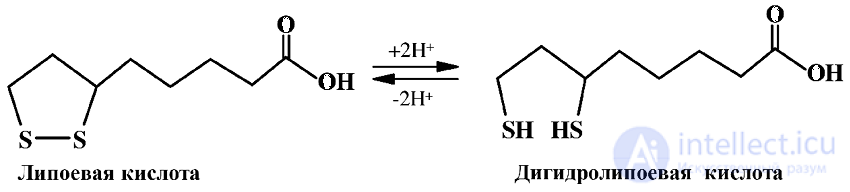
Lipoic acid exhibits biological activity as a prosthetic group of dehydrogenases, acting as a carrier of electrons and acyl groups. Lipoic acid and its amide bind to the enzyme through the amino group of lysine, with the disulfide bridge serving as the redox center.
In addition, lipoic acid is a powerful antioxidant and, together with ascorbic acid, acts as the main water-soluble antioxidant of the cell, protecting LDL from lipid peroxidation. The combined action of lipoic acid with vitamins E and C prevents the development of atherosclerosis.
Lipoic acid is conditionally essential for humans; the human body can synthesize it only in amounts sufficient to prevent its deficiency. The daily requirement for lipoic acid is approximately 1–2 mg. It is widely distributed in nature (plants, microorganisms), mainly in bound form but at low concentrations; the greatest amount is found in mitochondria and chloroplasts. The main dietary sources are yeast, meat products and milk.
Vitamin N plays an important role in cancer prevention and inhibits the degradation of insulin, hence its use in the treatment of diabetes mellitus. The main therapeutic form is lipoamide; both lipoamide and lipoic acid are produced as medicinal preparations and are used in the complex therapy of coronary atherosclerosis, liver diseases and various intoxications.
L-Carnitine (levocarnitine, vitamin B11, BT) is structurally an amino acid, γ-amino-β-hydroxybutanoic acid (more precisely, its trimethylated derivative). The name comes from the Latin word carnis — meat. Carnitine exists in two stereoisomeric forms, L-carnitine and D-carnitine; only L-carnitine exhibits biological activity.
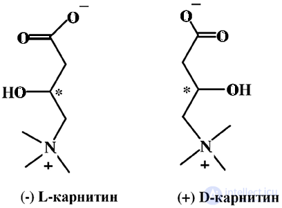
The D form has no effect on the body but acts as a competitive antagonist of L-carnitine (levocarnitine). Levocarnitine is a white crystalline powder, readily soluble in water and hot alcohol and practically insoluble in organic solvents. Carnitine occurs in animal muscle tissue, plants and microorganisms. Higher animals, including humans, synthesize levocarnitine from L-lysine with the participation of vitamins C, B3, B6, B9 and B12, iron and methionine.
Carnitine is one of the main sources of energy for muscle tissues. Its function in the body is associated with the transport of long-chain fatty acids, whose oxidation releases metabolic energy. Here L-carnitine acts as a coenzyme involved in the transport of fatty acids across membranes from the cytoplasm into the mitochondria. It interacts with the coenzyme (CoA) form of the fatty acid, forming a zwitterion — a derivative with a high affinity for the lipid layer of cell membranes. This ensures easy transport of the fatty acid across the mitochondrial membrane and then release of the acid as a result of hydrolysis:

Thus, carnitine intensifies the delivery of fatty acids into the cell and the formation of ATP, preventing fat deposition in the body, primarily in the heart, liver and skeletal muscle. Carnitine regulates the ratio of coenzyme A to acyl-coenzyme A, ensuring the operation of the tricarboxylic acid cycle and optimizing energy metabolism.
L-carnitine is synthesized in the liver and kidneys, from which it is transported to other tissues and organs. The level of L-carnitine is constant; it does not accumulate in the body, and excesses are effectively excreted. Carnitine is synthesized in the human body in large amounts (about 25% of the daily requirement) and is supplied with food (75%), so there is no convincing evidence of the need for its additional intake with food, although the use of meat broths rich in extractive substances to stimulate metabolic processes in debilitated patients is well known. Broth contains practically no proteins, fats or carbohydrates, but is rich in extractive substances, including carnitine, which increases the secretion and enzymatic activity of the gastric and intestinal digestive juices and improves the assimilation of food.
The recommended daily intake of carnitine is, mg: for adults — 300, for infants under one year — 10–15, from one to three years — 30–50, from four to six years — 60–90, from seven to eighteen years — 100–300. These intake norms increase several-fold in the presence of mental, physical or emotional overload.
The most carnitine is found in meat, poultry, seafood and dairy products. In grains, fruits and vegetables, carnitine is present in small amounts (Table 2.3.7).
Table 2.3.7. Carnitine content in foods
|
Food product |
Mass fraction, µg% |
|
Beef |
59,8–67,4 |
|
Chicken meat |
4,6–9,1 |
|
Heart |
19,3 |
|
Cow's milk |
0,53–3,91 |
|
Beef liver |
2,6 |
|
Beef kidneys |
1,8 |
|
Casein |
1,5 |
|
Avocado |
1,25 |
|
Wheat |
0,35–1,22 |
|
Peanuts |
0,76 |
|
Bread |
0,24 |
|
Cauliflower |
0,13 |
Levocarnitine is called the "growth vitamin" because it normalizes lipid and protein metabolism, reduces fat content in muscles and enhances metabolism, accelerating the build-up of muscle mass. The ability of carnitine to influence energy processes, reduce fatigue, increase physical endurance and reduce muscle pain after training by lowering the level of lactic acid in muscles is used in sports medicine. L-carnitine is used in pediatrics to increase resistance to psycho-emotional stress, in nervous and physical exhaustion, especially in elderly people, and in the postoperative recovery period. Many infant food manufacturers use it in the preparation of adapted milk-based and soy-based formulas.
At present there are several technologies for the industrial synthesis of L-carnitine: chemical synthesis, extraction from natural raw materials (from by-products of meat livestock farming) and microbiological synthesis (production of natural, 100% L-carnitine by microorganisms).
Methionine methylsulfonium chloride (S-methylmethionine, vitamin U). The name vitamin U comes from the Latin word ulcus — ulcer. Its structure is based on the amino acid methionine, whose sulfide group has been converted into a sulfonium group:
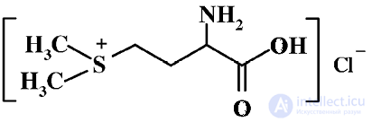
In nature, methionine methylsulfonium chloride is synthesized by many plants; asparagus, cabbage, celery, tomatoes, potatoes and tea leaves are the richest in it (Table 2.3.8). The main dietary source is the juice of raw vegetables; it is also present in fresh (unpasteurized) milk and in liver.
Table 2.3.8. Vitamin U content in foods (calculated on a dry weight basis)
|
Food product |
Mass fraction, mg% |
|
Asparagus, shoots |
100–160 |
|
White cabbage, leaves |
84,6 |
|
Unripe, green tomatoes |
46,7–48,2 |
|
White cabbage, core |
35,9 |
|
Ripe, red tomatoes |
18,7–47,5 |
|
Celery, stalk |
17,4 |
|
Table beet |
14,6 |
|
Parsley, greens |
6,4 |
|
Green tea |
4,6 |
|
Black tea |
– |
Methionine methylsulfonium chloride is readily soluble in water; it is destroyed when heated to 100 °C in a neutral and especially an alkaline medium, and is stable in an acidic one. Vitamin losses during technological processing of foods depend on the duration and intensity of heat exposure. Losses during blanching are 3–4%; with prolonged stewing the vitamin is completely destroyed.
Methionine methylsulfonium chloride is an active form of the essential amino acid methionine and acts as a donor of methyl groups in various biochemical reactions, mainly in methylation reactions with the formation of the fragments CH3OR, CH3NHR and CH3C, in the biosynthesis of choline, creatine and adrenaline.
In addition to methylmethionine sulfonium chloride, the bromide, iodide and sulfate forms have been isolated; they are used as antiulcer agents. Vitamin U is presumed to improve the metabolism of thiamine and choline, in particular in the gastric mucosa, and to increase its resistance to ulcer formation. Vitamin U increases the acidity of gastric juice and improves the motor function of the small and large intestine and of the gallbladder. It has lipolytic, antiallergic and antisclerotic effects.
The human body is unable to synthesize vitamin U and obtains it mainly from plant foods. The essentiality of S-methylmethionine has not been proven; in reactions involving the vitamin, when it is lacking in the human body, it is replaced by other substances. The daily requirement is approximately 500 mg.
Pangamic acid (vitamin B15, 6-O-N,N-dimethylglycyl-D-gluconic acid). The name comes from the Greek words pan — "everywhere" and gamy — "seed". Structurally it is a pentahydroxy-substituted hexanoic acid in which the hydroxyl group at C-6 is esterified with N,N-dimethylaminoacetic acid.
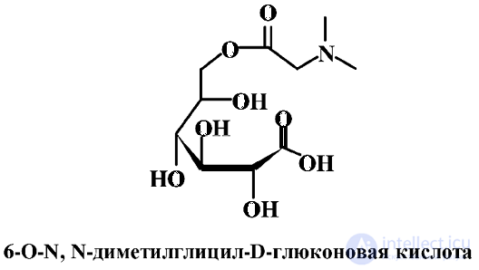
Pangamic acid was first isolated by E. Krebs from apricot kernels. It is widely represented in the seeds of plants — sesame, pumpkin, brown rice — and in the seeds of other cereal plants and the kernels of stone fruits; it is also present in brewer's yeast and liver.
The physiological action of pangamic acid consists in activating oxygen metabolism in the cells of various body tissues; in addition, it acts as a donor of methyl groups in methylation reactions. Pangamic acid has lipotropic properties and is necessary for normalizing lipid metabolism; it reduces the risk of liver disease. It takes part in the synthesis of creatine phosphate, which plays an important role in normalizing the functional capacity of muscles and improving energy processes in general. For this reason, pangamic acid deficiency causes diseases of the cardiovascular system, increased fatigue, premature aging and nervous disorders. In addition, pangamic acid removes toxins from the body, lowers serum cholesterol levels and reduces the craving for alcohol.
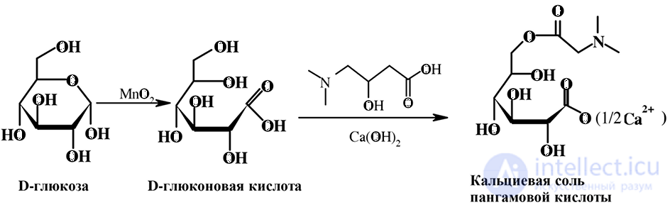
In medical practice, pangamic acid is used in the form of its calcium salt for the complex therapy and prevention of atherosclerosis, hepatitis, liver cirrhosis and alcohol intoxication. Pangamic acid is obtained by a chemical method: D-glucose is oxidized to D-gluconic acid, the primary hydroxyl group in it is then esterified with N,N-dimethylglycine, and the resulting ester is converted into the calcium salt:
The daily dose of pangamic acid for adults is 50–150 mg/day, and for children aged 3 to 14 years — 100–300 mg/day.

Comments