
Lecture
The human musculoskeletal system includes the skeletal (bony) and muscular systems. One of the leading functions of all living things, movement, is associated with its activity. There is no form of human activity that takes place without movement. In humans, the functions of the musculoskeletal system are linked to what gave humans an advantage over other representatives of the organic world: distinctly human qualities, labor and speech, which were the most important driving forces of anthropogenesis.
The most important functions of the skeleton are maintaining the shape of the body, protecting the internal organs, movement, hematopoiesis, and participation in mineral metabolism. The human skeleton consists of about 206 bones connected to one another by joints, ligaments, and other connections (Fig.).
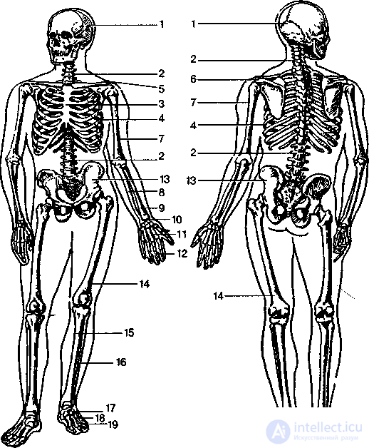
Fig. The human skeleton (anterior and posterior views):
1 – skull;
2 – vertebral column;
3 – sternum;
4 – thoracic cage;
5 – clavicle;
6 – scapula;
7 – humerus;
8 – ulna;
9 – radius;
10 – carpal bones;
11 – metacarpal bones;
12 – phalanges of the fingers;
13 – hip bone;
14 – femur;
15 – tibia;
16 – fibula;
17 – tarsal bones;
18 – metatarsal bones;
19 – phalanges of the toes.
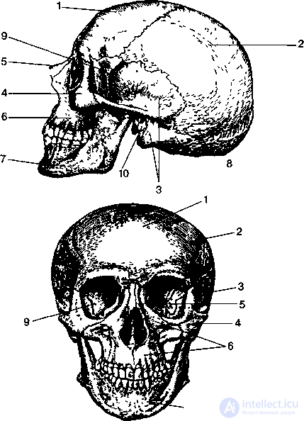
Fig. The skull, anterior and lateral views
frontal bone
parietal bone
temporal bone
zygomatic bone
nasal bone
maxilla
mandible
occipital bone
sphenoid bone
external acoustic opening
Bone is the main material from which the skeleton is built; it performs supportive, metabolic, and protective functions. In addition to bone tissue, bone contains blood vessels and nerves. The structural features of bone tissue determine the most important property of bone, its mechanical strength. For example, the tibia, part of the skeleton of the lower leg, when placed vertically, can withstand a load weighing almost two tons. The chemical composition of bones is also of great importance for their strength.
"... the external appearance of each bone is the realized idea of that bone's purpose." N.I. Pirogov (1843)
The human musculoskeletal system consists of a passive part (the skeleton and its joints) and an active part (the muscles). It performs the following functions:
As we have said, the human skeleton consists of bones (206) and the structures connecting them. The skeleton makes up up to 18% of body mass in men and about 16% in women.
In the course of phylogenesis, the skeleton passes through 3 stages of development:
Connective tissue (membranous);
Cartilaginous;
Bony.
Almost all bones of the skeleton pass through these three stages, with the exception of the bones of the cranial vault, some bones of the facial skeleton, and the clavicle.
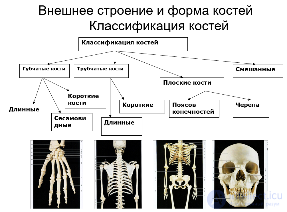
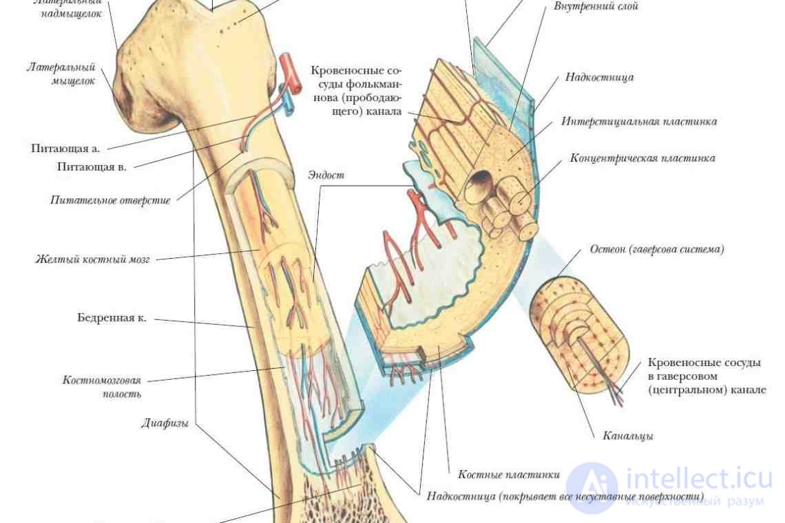
The bone tissue of an adult's skeleton contains mineral and organic substances in a ratio of 2:1. The former give bones hardness, the latter elasticity. The main organic component of bone is ossein. The inorganic compounds of bone are represented mainly by calcium salts, but bone tissue also contains sodium, magnesium, potassium, chlorine, fluorine, carbonates, and citrates in varying amounts. The chemical composition of bone is regulated by the hormones calcitonin and parathyroid hormone. The internal structure of bones is specially adapted to withstand deformation from compression and tension. On the outside, the bone is covered by a connective tissue membrane, the periosteum. In humans it is usually two-layered; the outer layer contains a plexus of blood vessels, which penetrate together with nerves into the interior of the bone. The inner layer of the periosteum contains collagen and elastic fibers and osteoblasts, actively dividing cells of bone tissue. Bundles of collagen fibers running from the periosteum form the basis for the attachment of tendons. The periosteum ensures the growth of bone in thickness and its regeneration after injury. Beneath the periosteum lies the compact bone. It is more developed in those bones whose main function is support and movement. Beneath the compact bone lies the spongy bone, which consists of a large number of bony trabeculae. They are arranged along the directions in which the bone experiences the pressure of gravity and the tension of the muscles attached to it. As a rule, the directions of the bony lamellae of two adjacent bones continue one another across the joint. In particular, in the complex of bones of the foot, the general direction of the bony lamellae has an arched shape. The cavities between the partitions of the spongy bone are filled with red bone marrow, which participates in hematopoiesis. The surface of many bones has roughenings, tubercles, and crests, the arrangement and degree of development of which are determined by motor loads. In men they are more pronounced than in women, and in people who engage in sports more than in those who do not (Fig. 59).
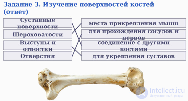
By shape, all bones are divided into four groups (Fig. 60): tubular (long and short), spongy (long, short, and sesamoid), flat, and mixed.
The long tubular bones include the femurs, humeri, and the bones of the forearm and lower leg. In them one distinguishes the middle part, the shaft, with a cavity inside filled in adults with yellow bone marrow, and the ends of the bones, covered with cartilage and forming the articular surfaces. The short tubular bones are found in the hand and foot.
The long spongy bones include the ribs and sternum, the short spongy bones include the vertebrae and the carpal and tarsal bones, and the sesamoid bones include the kneecap (patella).
Flat and broad bones are of small thickness but vary in size (scapula, parietal bones). Mixed bones differ in the diversity of their structure and combine spongy and flat elements (hip bones, mandible, zygomatic bones, occipital bone, etc.). Some mixed bones contain air-filled cavities (the temporal, maxillary, sphenoid, and ethmoid bones of the skull).
CHEMICAL COMPOSITION OF BONES
WATER – 50%
2. ORGANIC SUBSTANCES – 28.15% (protein OSSEIN)
3. INORGANIC SUBSTANCES – 21.85%:
The connections of the bones of the skeleton can be divided into three types: continuous, discontinuous (joints), and hemijoints (Fig. 61).
Continuous connections are characterized by almost complete immobility or slight mobility and are formed by connective tissue (sutures between bones, the attachment between a tooth and the wall of the tooth socket)
or cartilage (intervertebral discs, etc.). These connections are very strong and able to withstand considerable deformation.
Joints have the greatest mobility. Each joint consists of articular surfaces (at least two), an articular capsule, and a joint cavity. The joint cavity is a hermetically sealed space between the articular surfaces, separated from other organs by the walls of the articular capsule. The walls of the capsule consist of two layers: fibrous and synovial. The outer fibrous layer is formed of dense connective tissue and gives the articular capsule its strength. The inner synovial layer consists of a special tissue that produces the articular (synovial) fluid, which reduces friction between the articular surfaces and provides nutrition for the articular cartilage. Joints are reinforced by ligaments, most of which are derivatives of the outer layer of the articular capsule. Cylindrical, hinge, ellipsoid, saddle, and ball-and-socket forms of joints are distinguished.
In most cases the articular surfaces fit tightly against each other, which is ensured by the following factors:
negative pressure in the joint cavity relative to atmospheric pressure;
the tone of the muscles attached to the joint;
the shape of the articulating bones (the head of one corresponds to the socket of the other).
Excessive loads on a joint may result in its damage, sprain or rupture of ligaments, or displacement of the articulating ends of the bones (dislocation).
There is also a third (transitional) type of bone connection: hemijoints, or symphyses. Hemijoints lack an articular capsule, and the distinct cartilaginous layer between the bones has a cavity in its center filled with fluid similar in composition and properties to synovial fluid. Hemijoints have greater mobility than continuous connections. Example: the pubic symphysis of the hip bones.
The following main parts of the skeleton are distinguished:
The mass of the bones of an adult is about 18% of total body mass in men and 16% in women.
The skeleton of the head consists of the cranial and facial parts (Figs. 62, 63). The upper parts of the respiratory and digestive systems are attached to the bones of the skull. The brain is located inside the cranial part of the skull. This part has the shape of a rounded box and is formed by bones immovably connected to one another. It should be noted that the connections of the skull bones are mainly continuous and are made by means of sutures. There is only one discontinuous, movable joint, the temporomandibular joint, which enables raising and lowering of the mandible and its movements to the left, right, forward, and backward. In front, the cranial part of the skull contains the large unpaired frontal bone, on top the paired parietal bones, and on the sides the paired temporal bones. The unpaired sphenoid and ethmoid bones take part in forming the floor of the cranial part of the skull; the posterior wall is formed by the occipital bone, which has the large foramen magnum. The brain and spinal cord are joined through the foramen magnum. The circumference of the skull in an adult is 52–64 cm. The volume of the cranial cavity is about 1500 cm³.
The facial part of the skull includes: the maxillae (paired) and the mandible (unpaired), the nasal, zygomatic, lacrimal, and palatine bones, as well as the two inferior nasal conchae and the vomer, which take part in forming the walls of the nasal cavity. The facial bones also include the hyoid bone, to which the larynx is attached. Nerves and vessels pass through numerous canals and openings in the skull (they are located mainly in its lower part). The skull, mainly its facial part, houses the organs of hearing, vision, and smell, and also forms the skeleton of the nasal and oral cavities.
The skeleton of the trunk consists of the vertebral column and the thoracic cage. The vertebral column is a kind of axis of the body, whose upper end joins the skull and to whose lower end the bones of the pelvis are attached (Fig.). The vertebral column is formed of 33–34 vertebrae, each consisting of a body, an arch, and processes. Each vertebra has an opening inside, so that together they form the vertebral canal in the vertebral column, which contains the spinal cord. Five regions are distinguished in the vertebral column.
The size of the vertebral bodies increases from the cervical to the lumbar region because of the greater load on the lower vertebrae. The same reason, and
the attachment of the pelvic bones, leads to fusion and hypertrophy of the sacral vertebrae.
The vertebrae of the cervical, thoracic, and lumbar regions are connected by ligaments, joints, and cartilage. The latter are located between the vertebral bodies, are disc-shaped, and are formed of fibrocartilage, which has great strength and flexibility. The range of motion between two vertebrae is small, but overall these regions of the vertebral column have considerable mobility.
The human vertebral column has four functional curvatures. In the
cervical and lumbar regions the curvatures are convex forward and are called lordoses; in the thoracic and sacral regions they are convex backward and are called kyphoses. The curvatures of the vertebral column help absorb shock during walking, running, and jumping. In the course of postnatal ontogenesis, the cervical curvature forms first, when the child begins to hold up the head. The appearance of the lumbar and sacral curvatures is associated with upright walking.
The thoracic cage is formed by the sternum and 12 pairs of flat, arch-shaped ribs. Posteriorly, the heads of the ribs articulate movably (by means of joints) with the thoracic vertebrae, and anteriorly (except for the two lower pairs of ribs) they articulate with the sternum by means of their cartilages. The connections of the bones of the thoracic cage are quite mobile, which is important for breathing. The anteroposterior dimension of the thoracic cage is smaller than the lateral one. This shifts the body's center of gravity toward the vertebral column and increases stability during upright walking. Overall, the human thoracic cage has an egg-like shape, which changes somewhat depending on age, sex, occupation, and pathological influences.
It consists of the shoulder girdle and the free limb. The shoulder girdle is formed by the scapula, a flat triangular bone that articulates with the humerus by a ball-and-socket, highly mobile joint. The scapula articulates with the clavicle, the second bone of the shoulder girdle, by a plane, relatively immobile joint. With its other end the clavicle is movably joined to the sternum.
The free upper limb (or arm) consists of the upper arm, forearm, and hand. The upper arm is formed by a single tubular bone, the humerus. The forearm has two bones, the ulna and radius. The bones of the forearm together with the humerus form the complex elbow joint, and with the carpal bones the wrist joint. The hand includes eight small carpal bones arranged in two rows, five metacarpal bones forming the palm, and fourteen phalanges of the fingers, of which the thumb has two phalanges,
and the others three each. In humans the thumb is opposable to the other four fingers.
It is divided into the skeleton of the pelvic girdle and the skeleton of the free limb. The pelvic girdle includes the paired hip bones, each of which consists of three fused bones: the ilium (above), the ischium (below and behind), and the pubis (in front). The pelvic girdle together with the sacrum forms the pelvis,
which protects the internal organs of the abdominal cavity.
The free lower limb (leg) includes the thigh, lower leg, and foot. The thigh is represented by the long tubular femur. Its head at the upper end fits into the acetabulum of the hip bone, forming the ball-and-socket, triaxial hip joint, which is stronger but less mobile than the shoulder joint. The lower leg is formed by the tibia and fibula. The bones of the thigh and lower leg are joined by the knee joint, in which movements of flexion and extension of large amplitude take place. In front this joint is covered by a sesamoid bone, the kneecap (patella), which serves as a pulley over which the tendon of the quadriceps femoris muscle passes. The bones of the lower leg are joined with the tarsal bones by the hinge-type ankle joint. In the foot one distinguishes the tarsus, consisting of seven bones (the largest of which are the calcaneus and talus), the metatarsus, formed by five bones, and the phalanges of the toes. The big toe has two phalanges, the others three each. In connection with upright walking, the human foot has acquired the shape of an arch, which gives it spring-like properties and ensures a springy gait (Fig. 65).
In the course of prenatal and postnatal ontogenesis, a child's skeletal system undergoes complex transformations. The formation of the skeleton begins in the middle of the second month of embryogenesis and continues until 18–25 years after birth. At first, the embryo's entire skeleton consists of cartilaginous tissue, whose ossification is not complete by the time of birth, so a newborn's skeleton still contains a lot of cartilage. Even the bone itself differs significantly in chemical composition from the bone of an adult. In the first years it contains many organic substances, lacks strength, and bends easily under the influence of unfavorable external factors: narrow shoes, an incorrect position of the child in the crib, etc. Intensive thickening of the bones and an increase in their mechanical strength continue until 6–8 years. Then, until 14 years, the thickness of the compact layer of bone hardly changes, and during puberty an intensive increase in bone strength is again observed. Ossification of the skeleton is completed at 17–21 years in women and at 20–26 years in men. The bones of different regions ossify at different times. For example, ossification of the vertebral column is completed by 20–25 years, and that of the coccygeal vertebrae only by 30 years; ossification of the tubular bones of the hand is completed by 6–8 years, and of the carpal bones at 16–17 years. In this connection, strenuous fine manual work can disrupt the development of the bones of the hand, and wearing uncomfortable shoes can lead to deformities of the foot (most often to the development of flat feet). It should be noted that the rate of development of the bones of the hand correlates well with the general physical development of children and adolescents. Therefore, comparing chronological age with "bone" age gives a relatively accurate characterization of the rate of general physical development of children and adolescents and of their biological age.
Types of ossification (osteogenesis)
Age-related features of bones
|
|
Newborn |
Adult |
Old age |
|
Organic matter |
1/2 |
1/3 |
1/8 |
|
Inorganic matter |
1/2 |
2/3 |
7/8 |
With age, the porosity of bones increases, which leads to OSTEOPOROSIS (from Greek osteon – bone, poros – pore).
A newborn's vertebral column has no curvatures and is extremely flexible. By 3–4 years it acquires all four physiological curvatures. Cervical lordosis appears at 3 months, thoracic kyphosis at 6 months, and lumbar lordosis by 1 year. Sacral kyphosis forms last. However, until 12 years the child's vertebral column remains elastic and the curvatures are weakly fixed, which easily leads to abnormal curvatures under unfavorable developmental conditions. The most intensive growth of the vertebral column occurs at 7–9 years and during puberty. After 14–15 years the vertebral column hardly grows in length. The thoracic cage also approaches adult parameters by 12–13 years.
The pelvic bones fuse by 8–9 years, and at the same time its sex differences begin to form. Overall, the structure of the pelvis approaches the adult state by 14–17 years, and from this age the pelvis is able to withstand considerable loads.
The skull undergoes very large changes (Fig. 66). The fontanelles close at 1–2 years, and the fusion of the cranial sutures occurs only by four to five years. The facial part of the skull grows intensively during puberty until sexual maturity. The replacement of the primary teeth and the formation of the permanent teeth is completed by puberty, and only the third molars (wisdom teeth) appear after sexual maturation.
The timing of the eruption of the primary teeth and their replacement by permanent ones also correlates with general physical development and is used to determine the level of biological maturity of children and adolescents.
Thus, overall, the skeleton of children and adolescents is characterized by
high elasticity, which always poses a threat of its deformation
when hygiene standards are violated. An incorrect position of the child at
the desk, overloading of children and adolescents, and reduced motor
activity are risk factors for the development of skeletal pathologies.
The most common are various curvatures of the vertebral column (scoliosis,
that is, lateral curvatures).
Balanced, vitamin-rich nutrition is of particular importance for the proper development of the skeletal system. For example, with a deficiency of vitamin D, a disease called rickets may develop. It manifests as growth retardation and deformation of various parts of the skeleton: bowing of the legs, deformation of the skull, thoracic cage, and vertebral column.
Features of the newborn's skull
10. Presence of fontanelles, fonticuli:
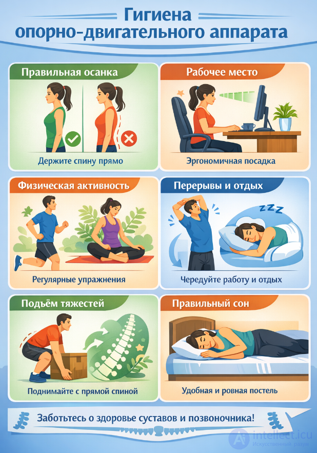
Hygiene of the musculoskeletal system is a system of rules and habits aimed at maintaining the health of bones, joints, and muscles, as well as preventing diseases and injuries. Posture, mobility, working capacity, and a person's general well-being depend on the condition of the musculoskeletal system.
The musculoskeletal system performs several important functions:
provides support for the body and shapes the organism;
participates in movement;
protects the internal organs;
participates in metabolism (for example, bones contain minerals: calcium and phosphorus).
Disorders in its function can lead to back pain, curvature of the spine, reduced mobility, and chronic diseases.
Posture is the habitual position of the body. Incorrect posture (slouching, forward head tilt) leads to overloading of the spine.
Important:
keep the back straight when walking and sitting;
do not lean too low over the desk;
distribute the load evenly on both sides of the body.
Especially important for schoolchildren and people who work at a computer:
the desk and chair should match the person's height;
the back should rest against the chair back;
the feet are on the floor;
the screen is at eye level.
Incorrect sitting causes muscle tension and can lead to curvature of the spine.
Regular exercise strengthens muscles and joints:
morning exercises;
sports (swimming, running, gymnastics);
walks in the fresh air.
Lack of movement weakens the muscles and worsens the condition of the joints.
Prolonged sitting or standing is harmful to the body.
Recommended:
take breaks every 30–60 minutes;
do light warm-up exercises;
change body position.
Improper lifting of heavy objects is a common cause of back injuries.
Rules:
lift the load with a straight back;
bend the knees;
keep the object close to the body.
For the health of bones and muscles, the following are necessary:
an adequate amount of calcium (dairy products);
vitamin D (sunlight, fish);
proteins (meat, legumes).
Poor nutrition can lead to weakening of bone tissue.
During sleep, the body recovers:
it is best to sleep on a moderately firm mattress;
the pillow should support the neck;
it is advisable to sleep 7–9 hours.
Observing hygiene helps to prevent:
curvature of the spine (scoliosis);
osteochondrosis;
joint diseases;
injuries and sprains.
Hygiene of the musculoskeletal system is the foundation of health and an active life. Simple daily habits, such as correct posture, physical activity and a well-organized workplace, help maintain the health of the spine and joints for many years. The earlier a person begins to follow these rules, the greater the chance of avoiding problems in the future.
Contractions of skeletal muscles provide movements of the body and maintain it in an upright position. Together with the skeleton, muscles give the body its shape. The activity of skeletal muscles is associated with the functions of respiration (work of the intercostal muscles and diaphragm), swallowing, hearing (work of the muscles that move the auditory ossicles), vision (movement of the eye in the orbit), vocal speech (work of the muscles of the larynx and tongue), etc.
The human body has about 600 skeletal muscles (Fig. 67). Muscle mass constitutes 35–45% of total body weight in men and 28–35% in women.
A skeletal muscle is an organ formed by striated muscle tissue that also contains connective tissue,
nerves (motor, sensory and autonomic) and vessels (blood and lymphatic). Each striated muscle (with the exception of the facial muscles) is enclosed in a connective tissue sheath (fascia) with a smooth surface, so it moves relative to neighboring muscles with minimal friction. Layers of loose connective tissue are also found inside the muscle, dividing the muscle fibers into separate groups (bundles). Moreover, each muscle fiber is covered with a thin layer of connective tissue. Blood vessels and nerves reach the muscle fibers within these connective tissue sheaths. The density of capillaries per unit area of muscle depends on its functional state. At its ends the muscle passes into a tendon (made of dense fibrous connective tissue), which has great strength but is unable to contract (Fig. 68). For example, the calcaneal (Achilles) tendon withstands a load of up to 300 kg. Tendon ends differ in shape (long, short, wide, fan-shaped), and the muscles are most often attached to different bones (Fig. 69). Skeletal muscles are classified by their relation to the joints (acting on one joint, two-joint, multi-joint), by their location in the human body (superficial, deep), and by the direction of the muscle fibers (circular or annular = sphincters, strap-like, fusiform, pennate). According to their functional significance, muscles can be divided into groups:
Muscles
by action on the joints
flexors / \ \ pronators extensors 1
by the activity performed
respiratory | facial expression masticatory
abductors
In carrying out any motor act, a whole group of muscles contracts. Muscles whose movements are combined, for example in flexion, are called synergists, or cooperating muscles, and muscles that take part in opposite actions are called antagonists. Antagonist muscles do not hinder the activity of synergist muscles: when the flexors contract, the extensors relax at the same time, which ensures coordination of movements. Muscles whose contraction moves a limb away from the body are called abductors, and their antagonists, which bring the limb closer to the body, are called adductors. Rotator muscles, when they contract, rotate a given part of the body (head, shoulder, forearm, etc.) toward the center (pronators) or away from the center (supinators).
, Contractility is the main property of muscles. It is characterized by the ability of a muscle to shorten or to develop muscle tension.
This ability of the muscle is related to the features of its structure and its functional properties.
Two types of muscle fibers are distinguished in skeletal muscles: slow (tonic) and fast (phasic). Some muscles contain only fast or only slow fibers, while others contain both at once. Thanks to the two types of fibers, the body can maintain posture and carry out movements.
The features of tonic muscles are as follows: they contain many mitochondria, and the source of energy is oxygen-dependent (aerobic) processes. In response to stimulation, a slow gradual contraction occurs, followed by slow relaxation, 100 times slower than that of fast fibers. Tonic muscles can contract for a long time, which ensures maintenance of posture. Tonic fibers are located in the deep layers of the muscles of the limbs and trunk.
Phasic fibers are characterized by a smaller number of mitochondria, so the main source of energy is anaerobic (oxygen-free) processes. These fibers respond to stimulation with rapid
contraction, but fatigue develops in them fairly quickly, as well as
oxygen debt. Phasic muscles are important for providing rapid movements. They are located closer to the surface of the body.
The activity of muscles is regulated by the central nervous system (CNS). Nerve impulses arising in various parts of the CNS eventually reach the motor neuron of the anterior horns of the spinal cord (motoneuron). Moreover, one motor neuron, as a rule, innervates several muscle fibers. It has been established that skeletal muscles contain about 250 million muscle fibers, whereas the number of motor neurons in the spinal cord is 420 thousand. Muscles of different parts of the body are innervated by different numbers of nerve cells. Thus, in the muscles of the eyeball one motor neuron innervates 3–6 muscle fibers, whereas in the leg muscles their number reaches 650. Thus, depending on the delicacy of motor acts and their biological significance, the number of neurons innervating muscles varies. A group of muscle fibers innervated by one motor neuron is called a motor unit. The large number of motor units can explain the smoothness of movements. If the nerve impulses reaching the muscle led to simultaneous excitation of all muscle fibers, movements would have a marionette-like, puppet-like character. But this does not happen, because impulses from different motor neurons reach the muscle not simultaneously but somewhat asynchronously. This is what promotes the gradual (graded) increase of contractions and the smoothness of the movements of our body.
Human movements, which are based on muscle contractions, are reflex in nature. The contractile mechanisms of muscle fibers are triggered by nerve impulses coming from nerve centers. The activity of the latter, in turn, is determined by stimuli coming from the environment through the activity of the sense organs. In addition, during the movement itself, the brain, on the basis of feedback (through a system of receptors located in the muscle itself, its tendons, or in the ligaments and joints), constantly receives signals about the progress of its execution. Thus a reflex ring is formed, which is a continuous flow of nerve impulses going from the peripheral receptors (proprioceptors) to the brain, from it to the effector organs (muscles), whose contractions are registered by the peripheral receptors, and from there the flow of nerve impulses again rushes to the nerve centers.
Any motor act, whether walking, running, or fine finger movements in writing or playing the piano, etc., involves delicate and precise coordination of the sequence of contractions of various muscle groups, their strength, and duration. Many parts of the CNS take part in the regulation of any movement. In the cerebral cortex, in the region of the precentral gyrus, lies the zone of the motor analyzer. The cortex carries out conditioned-reflex regulation of movements,
i.e., those movements that have been developed in a person through individual experience. Learning new forms of movement is possible only if the cortex is intact. The role of the diencephalon and its subcortical nuclei is that they regulate movements that have become automatic as a result of numerous repetitions. The cerebellum takes part in the regulation of unconditioned-reflex movements. Bilateral connections exist between the cortex, the cerebellum and the subcortical nuclei. At the level of the diencephalon, midbrain, medulla oblongata and spinal cord, reflex regulation of muscle tone is carried out.
Excitability and lability of muscles. In response to stimulation, a process of excitation develops in the muscle. This ability, as noted above, is called excitability. The level of muscle excitability is one of the most important indicators characterizing the functional state of the entire neuromuscular apparatus. The process of muscle excitation is accompanied by a change in metabolism in the muscle fibers, primarily a redistribution of K+ and Na+ ions between the intracellular and extracellular spaces.
The activity of a muscle is largely characterized by its lability — the speed or duration of the excitation process in excitable tissue. Muscle fibers have significantly lower lability than nerve fibers, but greater than the lability of synapses.
The levels of excitability and lability are not constant and change in different situations. Thus, light physical exertion (morning exercises) increases the excitability and lability of the neuromuscular apparatus, while significant physical and mental strain decreases them.
Tone of skeletal muscles. Even at rest, outside of work, muscles are not completely relaxed but are in a state of some steady involuntary tension (tone). This leads to a faster response to a stimulus and a stronger contraction. The external expression of tone is a certain degree of elasticity of the muscle. During mental and emotional strain the tone of various muscles may increase, and during deep sleep it decreases.
Isotonic and isometric muscle contraction. Contraction
of a muscle may be accompanied by its shortening, while the tension
remains constant. Such a contraction is called isotonic. If
the muscle tenses but no shortening occurs, the muscle contraction is called
isometric (for example, when trying to lift an immovable
load). Under natural conditions, muscle contractions are always
mixed in nature, and human movements are accompanied by both isotonic
and isometric muscle contractions. Therefore, one can speak
only of the relative predominance of isotonic or isometric
mode of muscle activity.
Under experimental conditions, a single nerve impulse is sufficient for muscle contraction. Such a muscle contraction is called a single twitch, and it proceeds very quickly, within several tens of milliseconds. Under natural conditions in the body, a series of impulses is always sent to the muscle. As a result, it does not have time to relax completely after the excitation caused by the previous impulse before a new impulse again causes its contraction, and so on. In other words, single contractions are summed into one longer contraction, which is called tetanic contraction, or tetanus. Its amplitude can be several times greater than the magnitude of the maximal single contraction. It is tetanus that ensures the duration and smoothness of the muscle contractions that occur under the natural conditions of our physical activity.
The magnitude of muscle contraction at a given strength of stimulation depends both on its structure and on the physiological state of the muscle fibers:
1. Long muscles contract by a greater amount than short ones.
2. The strength of a muscle depends on the number of muscle fibers in it: the greater the number of contracting fibers, the greater the contraction force developed by the muscle. Therefore, muscles with a pennate structure (containing a larger number of muscle fibers) are able to develop greater force than muscles with longitudinally arranged fibers.
3. Moderate stretching of a muscle increases its contraction. But with strong stretching, muscle contraction is weakened. The latter is due to the fact that the actin filaments lose their connections with the myosin filaments (they do not overlap) and the contractile apparatus of the fiber is unable to develop active force.
Working hypertrophy and atrophy of muscles. With systematic work of a muscle, the mass of muscle tissue increases. This phenomenon is called working hypertrophy of muscle tissue. It is based on an increase in the mass of cytoplasm and mitochondria and in the number of myofibrils, which leads to an increase in the diameter of the muscle fibers. In them, the processes of biosynthesis of nucleic acids, proteins, ATP and glycogen are accelerated. As a result, the strength and speed of muscle contraction increase. In the absence of load on the muscular system, in cases of prolonged bed rest, or in fractures, the opposite condition arises — muscle atrophy (hypotrophy).
A person's ability to perform physical (muscular) work for a long time is called physical work capacity. The magnitude of a person's physical work capacity depends on age, sex, training, environmental factors (temperature, time of day, oxygen content in the air, etc.) and the functional state of the body. For a comparative characterization of the physical work capacity of different people, the total amount of work performed in 1 minute is calculated and divided by body weight (kg), yielding relative physical work capacity (kg·m/min per 1 kg of body weight). On average, the level of physical work capacity of a 20-year-old young man is 15.5 kg·m/min per 1 kg of body weight, while in a young athlete of the same age it reaches 25. In recent years, determination of the level of physical work capacity has been widely used to assess the general physical development and health status of children and adolescents.
Prolonged and intense physical loads lead to a temporary decrease in the physical work capacity of the body. This physiological state is called fatigue. It has now been shown that the process of fatigue affects, first of all, the CNS, then the neuromuscular synapse, and last of all the muscle. The importance of the nervous system in the development of fatigue in the body was first noted by I.M. Sechenov. The fact that interesting work does not cause fatigue for a long time, while uninteresting work does so very quickly, can be regarded as evidence of the validity of this conclusion, even though the muscular load in the first case may even exceed the work performed by the same person in the second case.
Fatigue is a normal physiological process, developed through evolution to protect the body's systems from systematic overwork, which is a pathological process and is characterized by disruption of the activity of the nervous system and other physiological systems of the body.
During ontogenesis, the muscular system undergoes significant
structural and functional changes. The formation of muscle cells
and the formation of muscles as structural units of the muscular system occurs
heterochronically, i.e., first those skeletal muscles form that
are necessary for the normal vital functions of the body
at the given age stage. The process of "rough" formation
of muscles ends by week 7–8 of prenatal development. After birth
the process of formation of the muscular system continues. In particular, intensive growth of muscle fibers is observed up to 7 years of age and during puberty. By 14–16 years the microstructure of skeletal muscle tissue is almost fully mature, but thickening of muscle fibers (improvement of their contractile apparatus) can continue until 30–35 years.
The development of the muscles of the upper limbs outpaces the development of the muscles of the lower limbs. In a one-year-old child, the muscles of the shoulder girdle and arms are developed considerably better than the muscles of the pelvis and legs. Larger muscles always form earlier than smaller ones. For example, the forearm muscles form earlier than the small muscles of the hand. The muscles of the arms develop especially intensively at 6–7 years. The total muscle mass increases very rapidly during puberty: in boys at 13–14 years, and in girls at 11–12 years. Below are data characterizing the mass of skeletal muscles during postnatal ontogenesis.
Age
0–10 days
8 years
12 years
15 years
18 years
Muscle mass, % of total body weight
23.3 27.2 29.4 32.6 44.2
The functional properties of muscles also change considerably during ontogenesis. The excitability and lability of muscle tissue increase. Muscle tone changes. A newborn has increased muscle tone, and the flexor muscles of the limbs predominate over the extensor muscles. As a result, the arms and legs of infants are more often in a flexed position. Their ability of muscles to relax is poorly expressed (this is the cause of some stiffness in children's movements), and it improves with age. Only after 13–15 years do movements become more fluid. It is at this age that the formation of all parts of the motor analyzer is completed.
During the development of the musculoskeletal system, the motor qualities of muscles change: speed, strength, agility and endurance. Their development is uneven. First of all, speed and agility develop.
Speed (rapidity) of movements is characterized by the number of movements that a child is able to perform per unit of time. It is determined by three indicators:
the speed of a single movement,
the motor reaction time, and
the frequency of movements.
The speed of a single movement increases considerably in children from 4–5 years and reaches the adult level by 13–15 years. By the same age the simple motor reaction time, which is determined by the speed of physiological processes in the neuromuscular apparatus, also reaches the adult level. The maximal voluntary frequency of movements increases from 7 to 13 years, and in boys at 7–10 years it is higher than in girls, while from 13–14 years the frequency of movements in girls exceeds that in boys. Finally, the maximal frequency of movements in a given rhythm also increases sharply at 7–9 years. On the whole, the speed of movements develops maximally by 16–17 years.
By 13–14 years the development of agility is mostly completed; agility is related to the ability of children and adolescents to perform precise, coordinated movements. Consequently, agility is related to:
the spatial accuracy of movements,
the temporal accuracy of movements,
the speed of solving complex motor tasks.
The preschool and early school periods are most important for the development of agility. The greatest increase in the accuracy of movements is observed from 4–5 to 7–8 years. Interestingly, athletic training has a beneficial effect on the development of agility, and in 15–16-year-old athletes the accuracy of movements is twice as high as in untrained adolescents of the same age. Thus, until 6–7 years children are not able to perform fine, precise movements in an extremely short time. Then spatial accuracy of movements gradually develops, followed by temporal accuracy. Finally, last of all, the ability to quickly solve motor tasks in various situations improves. Agility continues to improve until 17–18 years.
The greatest increase in strength is observed in middle and senior school age; strength increases especially intensively from 10–12 years to 16–17 years. In girls the increase in strength begins somewhat earlier, from 10–12 years, and in boys from 13–14 years. Nevertheless, boys surpass girls on this indicator in all age groups.
Endurance develops later than the other motor qualities; it is characterized by the length of time during which a sufficient level of the body's work capacity is maintained. There are age-related, sex-related and individual differences in endurance. The endurance of preschool children is at a low level, especially for static work. An intensive increase in endurance for dynamic work is observed from 11–12 years. Thus, if the volume of dynamic work of 7-year-old children is taken as 100%, then in 10-year-olds it will be 150%, and in 14–15-year-olds more than 400%. Endurance for static loads also increases just as intensively in children from 11–12 years; by 17–19 years endurance is about 85% of the adult level. It reaches its maximum level by 25–30 years.
The development of movements and of the mechanisms of their coordination proceeds most intensively in the first years of life and in adolescence. In a newborn, coordination of movements is very imperfect, and the movements themselves have only an unconditioned-reflex basis. Of particular interest is the swimming reflex, whose maximal manifestation is observed at about the 40th day after birth. At this age a child is able to make swimming movements in water and stay afloat for up to 15 minutes. Naturally, the child's head must be supported, since the child's own neck muscles are still very weak. Subsequently the swimming reflex and other unconditioned reflexes gradually fade, and motor skills form to replace them. All the basic natural movements characteristic of humans (walking, climbing, running, jumping, etc.) and their coordination are formed in a child mainly by 3–5 years. The first weeks of life are of great importance for the normal development of movements. Naturally, coordination mechanisms are still very imperfect in preschool age as well. Despite this, children are able to master relatively complex movements. In particular, it is at this age that they learn tool-use movements, i.e., motor abilities and skills of using an instrument (a hammer, a key, scissors). From 6–7 years children master writing and other movements requiring fine coordination. By the beginning of adolescence the formation of coordination mechanisms is generally completed, and all types of movements become accessible to adolescents. Of course, improvement of movements and their coordination through systematic exercise is possible in adulthood as well (for example, in athletes, musicians, etc.).
The improvement of movements is always closely connected with the development of the child's nervous system. In adolescence, coordination of movements is very often somewhat impaired owing to hormonal changes. Usually by 15–16 years this temporary deterioration disappears without a trace. The general formation of coordination mechanisms is completed at the end of adolescence, and by 18–25 years they fully reach the level of an adult. The age of 18–30 years is considered the "golden" age in the development of human motor function. This is the age of the flowering of a person's motor abilities.

Comments