
Lecture
Chemical substances enter the body through the respiratory tract, the gastrointestinal tract and through the skin. The behavior of foreign compounds in the body is presented in Diagram 4.
Diagram 4.
Behavior of poisons in the body
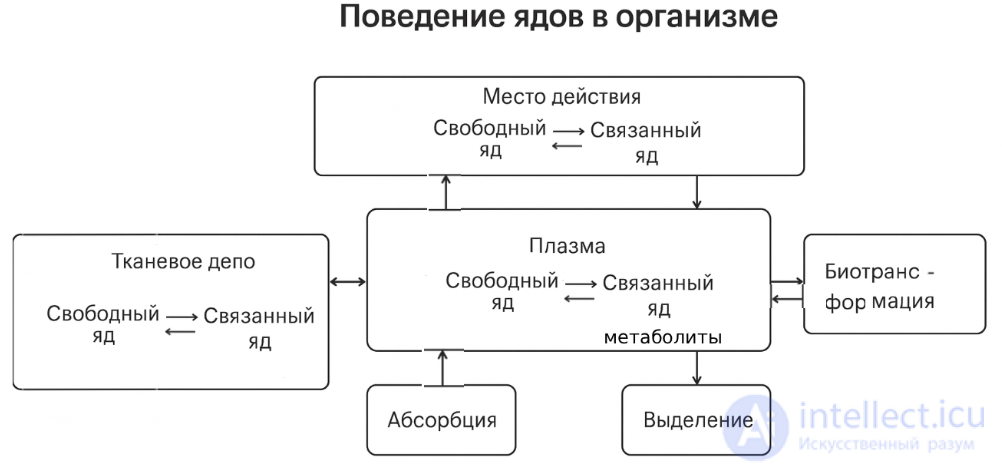
However, many industrial poisons pass through a number of biological membranes of the body and have a shorter path, usually determined by their physicochemical properties.
Inhalation entry of harmful substances. The rate of entry of harmful substances into the body through the respiratory tract is determined, first of all, by their physical state (gases, mists, dust, fumes).
Inhalation poisonings are characterized by the most rapid entry of the poison into the blood. This is explained by the large surface area of the pulmonary alveoli (100 –150 m2), the small thickness of the alveolar membranes, the intense blood flow through the pulmonary capillaries, and the absence of conditions for deposition of the poison. Absorption of volatile compounds already begins in the upper respiratory tract, but it takes place more completely in the lungs and proceeds according to the law of simple diffusion. Depending on the chemical activity of gases, their ability to penetrate and act on the body varies.
A distinction is made between non-reacting electrolytes, which do not change their composition upon entry into the body, and reacting ones, which undergo transformations. Non-reacting electrolytes include organic substances, for example alcohols, acetone, ethers, hydrocarbons, halogenated hydrocarbons, and others. Reacting compounds include vapors of inorganic acids.
The rate and extent of retention of gases mainly depend on their physicochemical properties. The retention of toxic substances in the respiratory tract must be linked to their simultaneous entry into the blood and tissues of the body.
When a constant concentration of a harmful substance is inhaled, the content of the poison in the blood first increases rapidly and then stabilizes at approximately the same level. The content of the poison in venous blood gradually equalizes with its concentration in arterial blood. This occurs because the body gradually becomes saturated with the poison, and its absorption slows down substantially.
The main physicochemical indicator determining the absorption of a poison into the blood is the coefficient of solubility of vapors in water (the Ostwald coefficient):
|
water |
|
|
= ----------- , |
(6) |
|
air |
|
It characterizes the distribution of volatile compounds between the liquid and gaseous phases on reaching equilibrium. The higher this coefficient, the more of the substance passes from air into the blood. The solubility coefficient also determines the rate at which equilibrium is established between the content of the harmful substance in the air and in the blood. Non-electrolytes with a high solubility coefficient (ethyl alcohol, acetone) pass slowly from air into the blood. Compounds with a low solubility coefficient (hydrocarbons) quickly reach an equilibrium concentration between arterial blood and alveolar air, since they not only dissolve in the liquid part of the blood, but are also able to bind to plasma proteins.
The basic physiological indicators of the body - the intensity of respiration and blood circulation - depend on the severity of the work being performed. When poisons enter a body that is in a stressed state, the rate of reaching equilibrium in the blood/air system increases, especially for substances with a relatively high solubility coefficient. An increase in the rate of blood circulation in the body primarily affects the retention of chemical compounds having solubility coefficients less than unity.
It has been established that at equilibrium, humans retain in the body from 40 to 65% of benzene, from 41 to 63% of toluene, from 46 to 96% of trichloroethylene, and of chloroform (under anesthesia) first from 74 to 80%, and then about 60%.
When reacting vapors of inorganic acids enter the body, their retention occurs at a more constant rate than that of volatile non-electrolytes. Reacting vapors and gases have the ability to damage the alveolar membrane itself, disrupting its barrier and transport function, which leads to the development of toxic pulmonary edema.
Particles of solid and liquid substances form aerosols in the air of the work zone. When aerosols enter the body through the respiratory tract, particles settle along its entire length. The degree of penetration of an aerosol into the body depends on the size and shape of the particles, their charge, etc. Relatively large particles usually settle at points where air flows change direction, for example, when a particle strikes the wall of the respiratory tract. Smaller particles settle in the lower part of the respiratory tract. Particles larger than 10 µm settle completely in the nasopharynx, particles with a diameter greater than 2 µm and less than 10 µm are retained in the upper respiratory tract, while particles smaller than 2 µm settle in the alveolar region.
In the region of self-cleaning of the respiratory tract, particles that have settled on the mucous membrane of the upper
part of it move upward together with the mucus and are gradually removed from the body. However, when water-soluble toxic aerosols enter the body, resorption of the poison occurs (that is, the ability to cause a toxicological effect). In this case, poisoning occurs along the entire length of the respiratory tract.
From a hygienic point of view, aerosols are divided into: toxic, fibrogenic and inert. A very important property of aerosols upon entry into the body is their dispersity. The size of solid or liquid particles determines the length of time they remain in the air of the work zone and the depth of penetration into the respiratory tract. Aerosols can be formed by dispersion and condensation. When solid particles are crushed, dispersed aerosols are formed. Moreover, the harder the substance being ground, the smaller the size of the resulting particles. Aerosols are formed from vapors of metals and their compounds by condensation, for example during welding. An aerosol, having penetrated into the alveolar space of the lungs of the body, either dissolves in the blood of the blood vessels or is retained in the alveoli. Depending on the properties of the solid particles (aggressive or inert), they are removed from the body with the mucus or
penetrate into the tissue of the alveoli, which degenerates, forming scar tissue and becoming impermeable to air. This process, occurring in the tissues of the lungs, is irreversible. Dusts that cause such scar growths in the lungs are called fibrogenic or pneumoconiosis-hazardous, and the diseases – pneumoconioses.
Fibrogenic dusts are divided into two main groups: mineral and organic, and each of them is in turn subdivided into dusts: quartz-containing, inert and fibrous.
Quartz-containing dust contains two percent or more of free silicon dioxide. Dusts of mineral and organic origin with a lower content or complete absence of silicon dioxide are inert and do not possess fibrogenic and toxic properties. Dusts of mineral and organic origin (asbestos, cotton, etc.) whose fiber length exceeds their diameter by tens of times are classified as fibrous.
The most dangerous form of pneumoconiosis is silicosis - an occupational disease caused by exposure to dust with a high percentage content of silicon dioxide. It has been established that the mass or surface area of silicogenic dust is of great importance in the development of silicosis. Dust particles measuring 1-5 µm are the most silicosis-hazardous.
The presence in the air of fibers of various types of asbestos is of particular importance, since their entry into the body leads to the formation of malignant tumors. It has been established that the greatest influence on their formation is exerted by asbestos fibers with a diameter of less than 0,25 µm and a length of more than 8 µm, which is why it is classified as a carcinogenic substance.
Dust from synthetic fibers (nylon, polyester, etc.) aggravates chronic diseases of the respiratory organs and skin. These include: chronic bronchitis, pneumonia, rhinitis, laryngitis, dermatitis and eczema.
The biological action of polymer dust is explained by the content of residual monomers in them; for example, for nylon this is caprolactam, for nitron – acrylonitrile, and for polyester – dimethyl terephthalate.
The effect of inert aerosols on the lungs also does not pass without a trace, however, no change in alveolar volume occurs and no scar tissue forms in the lungs in this case. These
effects on the body are reversible in nature, since inert aerosols are removed from the lungs. However, at a concentration exceeding their MAC (more than 10 mg/m3), irritation of the mucous membranes, eyes and skin occurs. The solubility of aerosols in water (tissue fluids and blood) has both a positive and a negative value. If the dust is non-toxic and its action on lung tissue is limited to mechanical irritation, then good solubility of the aerosols promotes their removal from the lungs. In the case where the dust is toxic, its good solubility is a negative factor, since it causes poisoning of the body.
When harmful substances enter the body orally, their entry into the blood can occur in various ways. The entry of toxic substances through the mouth under industrial conditions can occur through swallowing dust, smoking, eating, and so on. A number of poisonous fat-soluble compounds, which include phenols, certain salts and especially cyanides, are absorbed into the blood in the oral cavity, due to diffusion from the oral cavity through the mucous membrane. This leads to an increase in the toxicity of the poison, since it enters the liver bypassing the stomach.
The remaining substances enter the gastrointestinal tract (GIT) and are subjected to the decomposing action of gastrointestinal juices. Throughout the GIT there are significant gradients of acid-base reaction, which determine the varying rate of absorption of toxic substances. The acidity of gastric juice is close to unity, therefore all acids in the GIT are in a non-ionized state and are easily absorbed. Toxic substances
in the stomach can be sorbed by food masses, be diluted by them, as a result of which contact of the poison with the mucous membrane decreases. In addition, the rate of absorption is affected by the intensity of blood circulation in the mucous membrane of the stomach.
The absorption of poisonous substances mainly takes place in the small intestine. Some substances, for example heavy metals, directly damage the intestinal epithelium and disrupt absorption. In the intestine, as in the stomach, fat-soluble substances, as well as electrolytes, are absorbed by diffusion. However, the absorption of electrolytes depends on their ionization. This determines the rapid resorption of bases, for example aniline.
Strong acids and bases are absorbed slowly, forming complexes with intestinal mucus. Metals are absorbed mainly in the upper part of the small intestine; chromium, manganese and zinc – in the ileum, iron, copper, mercury, antimony – in the small intestine. Alkali metals are resorbed
quickly and completely, while alkaline-earth metals are absorbed in an amount of 20-60%, since they form poorly soluble complexes with phosphates and fatty acids. Protein complexes of rare-earth metals are absorbed with particular difficulty. Upon absorption from the stomach and intestine, substances primarily enter the liver, where various transformations of many poisons take place. Mainly, these transformations are aimed at detoxifying the poisons, however, another process is also possible – the formation of more hazardous compounds (the so-called lethal synthesis).
The slowing of regional blood flow and the deposition of venous blood in the intestinal region during a number of severe poisonings accompanied by shock promotes the creation of a depot of toxic substances in the GIT. To reduce poisoning, its thorough cleansing is necessary not only at early but also at later stages.
Penetration of harmful substances through the skin. This type of toxin penetration is called percutaneous. Gaseous, liquid and solid substances, belonging predominantly to non-electrolytes, can penetrate through the skin. Heavy metals and their salts are absorbed into the blood through the skin barrier to an insignificant degree. Among industrial poisons causing intoxication upon penetration through the skin, first place is held by aromatic nitro- and amino compounds, organophosphorus pesticides, certain chlorinated hydrocarbons and organometallic compounds.
There are three possible routes for the penetration of various substances through the skin: through the epidermis, through the hair follicles, and through the excretory ducts of the sebaceous glands.
The main barrier to the penetration of harmful substances through the skin is formed by the various layers of the epidermis (the upper stratum corneum and the stratum lucidum). It should be taken into account that the salts of many metals, combining with fatty acids and skin sebum, can be converted into fat-soluble compounds and penetrate through the barrier layer of the epidermis. Lead, tin, copper, arsenic, bismuth, antimony, mercury and thallium are absorbed into the blood better than other metals. Zinc and cadmium form protein complexes and also penetrate through the skin. Hexavalent chromium, upon penetrating through the skin, is reduced to the trivalent state, which possesses increased toxicity.
Mechanical damage to the skin (abrasions, scratches and wounds), thermal and chemical burns promote the penetration of toxic substances into the body.
The general spatial diagram of the movement of the poison in the body is presented in Diagram 5.
Diagram 5.
Routes of entry of the poison into the body and its elimination into the external environment (according to E.A. Luzhnikov, 1994).
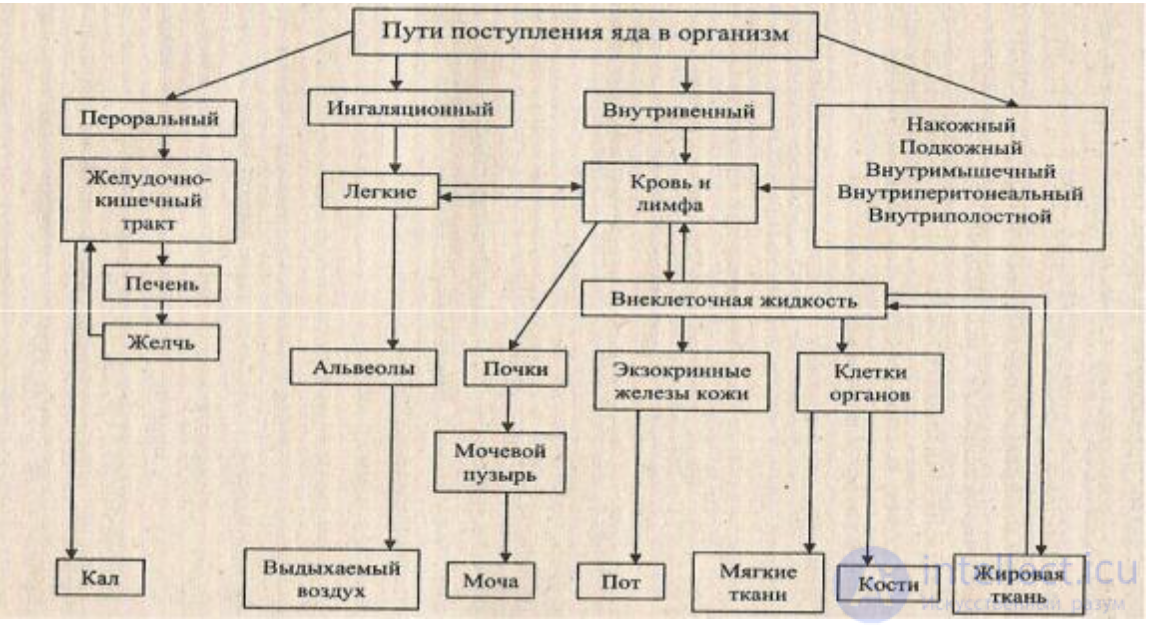
The entry of poisons into the body, their distribution among organs and tissues, metabolism and excretion from the body are largely determined by their ability to pass through biological membranes and by the character of the interaction of harmful substances with them.
Whether or not poisoning develops following the entry of a poison into the body, what degree it will reach, and how long it will last depend to a great extent on what transformations will occur with the poison in the body, and at what rate. All these processes are called the fate of the poison
in the body. From the moment of its entry until it reacts with its point of application (the receptor), the poison is subject to the action of a variety of biological factors. It is carried by the bloodstream throughout the whole body, penetrates into tissues and organs through the capillary network, is sometimes deposited in some of them (for example, lead in bones), undergoes various transformations and, finally, is excreted from the body unchanged or in the form of metabolites. Transformation of the poison may already occur at the site of contact with tissues. In the blood, some toxic compounds bind to plasma proteins, mainly albumins. This reduces the possibility of the poison interacting with receptors or slows this process down. Thus, the processes of the entry of poisons into the body, their distribution and transformation, take place over time. The study of such time dependences in the body is the subject of toxicokinetics. These studies make it possible to more effectively establish the maximum allowable levels (MAC) of substances in water, air, soil and food products.
The distribution of toxic substances in the body depends on 3 main factors (Diagram 6):
Diagram 6.
Main factors determining the development of acute poisoning.
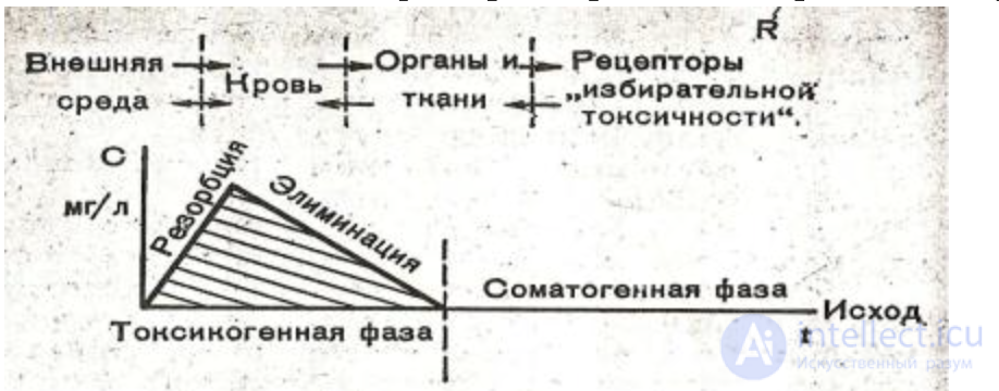
R – spatial; C – concentration; t – temporal. Main pathological syndromes of the toxicogenic phase: shock, asphyxia, coma, hemorrhage; somatogenic phase: pneumonia, acute renal failure, acute hepatic-renal failure, trophic disorders, sepsis.
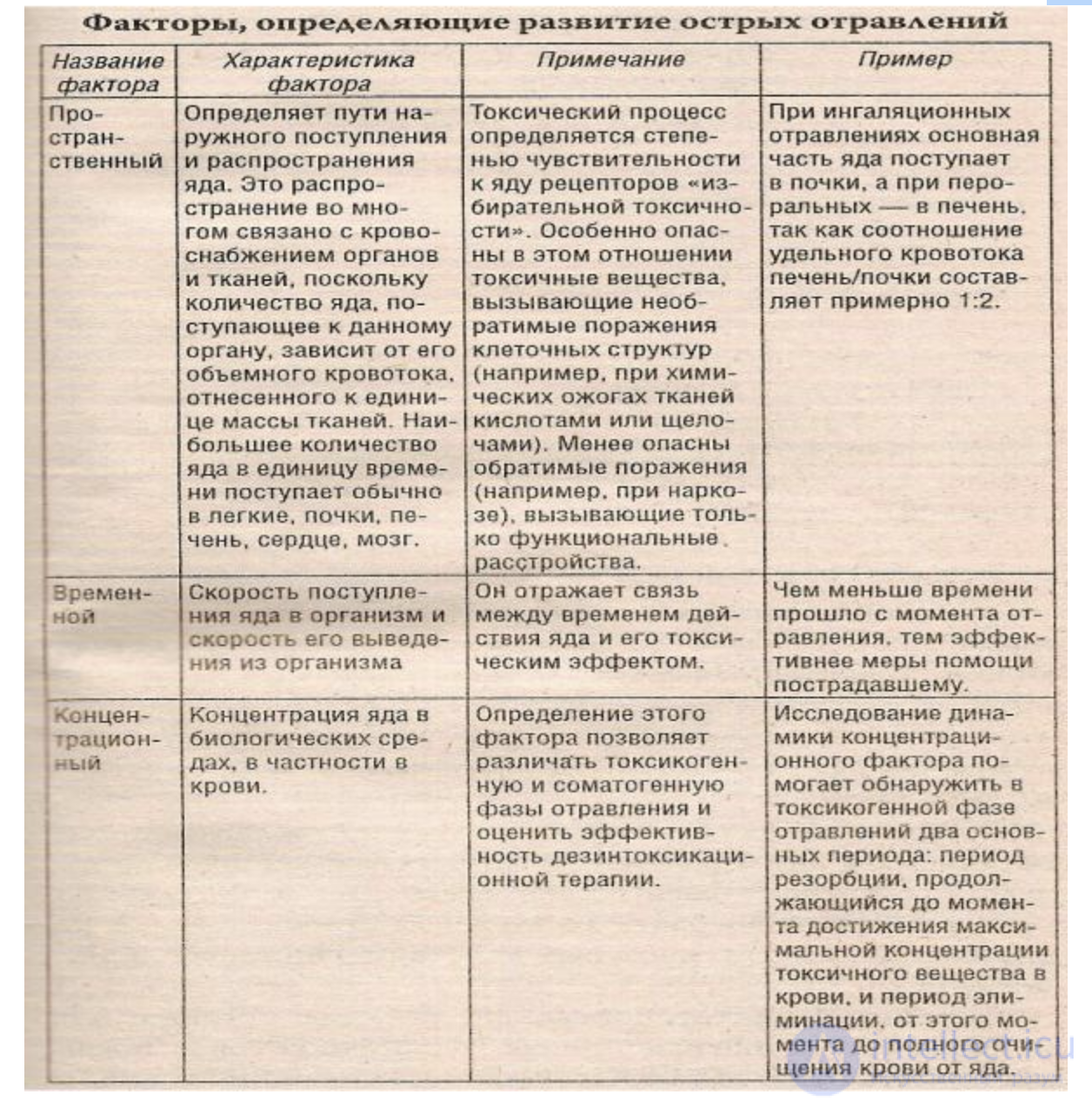
1. The spatial factor determines the routes of external entry and distribution of the poison.
2. The temporal factor determines the rate of entry into the body and the rate of its elimination from the body.
3. The concentration factor, i.e., the concentration of the poison in biological media, is considered the main factor in clinical toxicology. Determination of this factor makes it possible to distinguish between the toxicogenic and somatogenic phases of poisoning and to assess the effectiveness of detoxification therapy. Study of the dynamics of this factor helps to identify, within the toxicogenic phase of poisoning, two main periods: the resorption period, lasting until the moment the maximum concentration of the toxic substance in the blood is reached, and the elimination period, from that moment until the blood is completely cleared of the poison.
Diagram 7 presents the stages of acute poisoning and the factors determining their development.
Diagram 7.
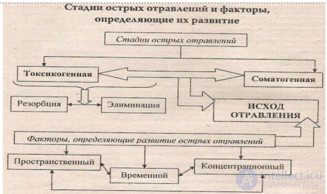
Table 5 gives the main characteristics of the poisoning factors.
Table 5.
According to the hypothesis of the well-known toxicologist E. Albert, any chemical substance, in order to produce a biological effect, must possess at least two independent characteristics:
1. Affinity for receptors.
2. Its own physicochemical activity.
The entry of foreign substances into the body, their distribution among organs and tissues, biotransformation and excretion presuppose their penetration through a number of biological membranes. The membrane system of the body has the same structure but differs in its functional properties.
The most important condition for the existence of the cell and, consequently, for life is the normal functioning of biological membranes. The membrane is an extremely thin (6-10 nm) and fairly dense film covering the entire cell (Fig. 3).
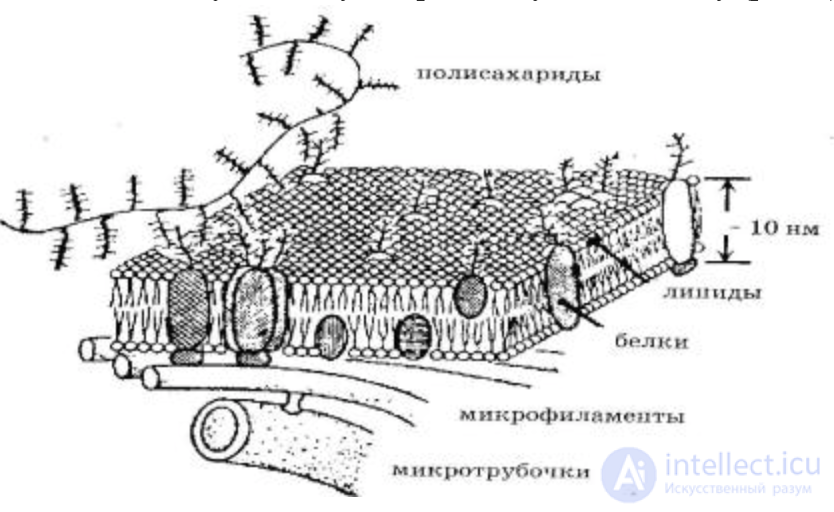
Fig. 3. Structure of the biology of the biological membrane of the cell
The main chemical compounds forming the membrane are the orderly arranged molecules of phospholipids and proteins. A phospholipid molecule contains a polar part (a derivative of phosphoric acid) and a long nonpolar part (a tail consisting of fatty acid residues). In the polar part of the phospholipid molecule there are two charged groups located at some distance from each other, which form an electric dipole. Membranes contain various phospholipids; for example, there are about 20 types of them in the erythrocyte membrane. They may be polar, neutral, or have an uncompensated negative charge. The hydrocarbon tails of the phospholipid molecule contain approximately 20 carbon atoms, and the tail itself may have 1-4 double unsaturated bonds. Phospholipid and protein molecules are held together by noncovalent interactions.
The inner layer of the membrane consists of two rows of lipids. They are esters of fatty acids and an alcohol. The lipid molecules are arranged so that their nonpolar hydrophobic ends face the internal and external environment. The lipid layer is not continuous. In certain places the membrane is penetrated by protein molecules, forming hydrophilic pores in them, through which water-soluble substances pass. The protein pores are anchored by cytoplasmic structures of the cell. These include microfilaments and microtubules (Fig. 3). Microtubules are hollow cylinders about 300 nm in diameter with a wall thickness of 5 nm, built from a special protein (tubulin). Microfilaments are thin filaments found throughout the cytoplasm of cells. They are especially numerous in the surface layer of the cytoplasm, where they form a dense network of thin filaments crossing in different directions. Microfilaments are formed from the protein actin, whose molecules form a long fibril consisting of two helices twisted relative to one another. Actin microfilaments interact with microtubules and provide the motor activity of the cell.
In addition to phospholipids and proteins, biological membranes contain other chemical compounds as well. Animal cell membranes contain a large amount of cholesterol, and also contain glycolipids and glycoproteins, which belong to steroids (lipids). On the surface of the membranes of all cells there are carbohydrates, which are linked to proteins by a covalent bond. The carbohydrate content in membranes ranges from 2 to 10% by mass. The polysaccharide layer has a thickness of 10 to 20 nm.
All cell membranes are mobile, fluid structures: most of the molecules of proteins, lipids, polysaccharides, water, and ions of potassium, sodium, calcium, etc. that make them up are able to move fairly quickly within the plane of the membrane, changing their position within it. In this case, the migration of substances occurs both by diffusion and actively, with the consumption of energy. Membranes are a dynamic system and therefore quickly recover after damage; moreover, they are able to stretch and contract during cell movements.
The membranes of different types of cells differ substantially both in their chemical composition and in the relative content of proteins, glycoproteins, and lipids in them, and consequently in the nature of the receptors present in them. Each type of cell is therefore characterized by an individuality determined by the presence of glycoproteins, whose branched chains take part in recognizing environmental factors and in the response of cells to their action. This phenomenon is observed in the process of tissue differentiation. In this case, cells that are similar in structure are correctly oriented relative to one another with the help of the recognition sites of the membrane, thereby ensuring their adhesion and the formation of tissues. Recognition is also linked to the regulation of the transport of molecules and ions across the membrane, as well as to the immune response, in which glycoproteins play the role of antigens. Membranes also contain specific receptors, electron carriers, energy converters, and enzymatic proteins. Proteins take part in ensuring the transport of certain molecules into or out of the cell, provide the connection of the cell skeleton with the cell membranes, and also serve as receptors for receiving and converting chemical signals from the environment.
This structural organization and orderliness of the biological membrane is responsible for such a vital function as semipermeability, that is, the ability to selectively let various molecules and ions into and out of the cell. Owing to this, the appropriate concentration of ions is created and maintained in the cell, and osmotic processes take place.
One of the most important functions of the biological membrane is the generation and transmission of biopotentials. This phenomenon underlies the excitability of cells, the regulation of intracellular processes, the functioning of the nervous system, and muscle contraction. Diagnostic methods, for example electrocardiography, are based on the study of the electric fields created by the biopotentials of organs and tissues.
In the course of vital activity, differences in electric potential of various origins may arise in cells and tissues:
1) redox potentials are formed by the transfer of electrons from some molecules to others;
2) membrane potentials - are formed when a concentration gradient of ions arises and they are transported through
the membrane.
The membrane potential is the potential difference between the internal (cytoplasmic) and external surfaces of the membrane:
M = IN - OUT (7)
Membrane potentials are subdivided into resting potentials and action potentials.
The resting potential is the difference in electric potentials between the internal and external surfaces of the membrane in the unexcited state. It is determined by the different concentration of ions on either side of the membrane and by the diffusion of ions.
If the concentration of some ion inside the cell CIN differs from the concentration of that ion outside COUT and the membrane is permeable to that ion, a flow of particles through the membrane arises. This disrupts the neutrality of the system, and a potential difference is formed inside and outside the cell, which will impede the further movement of ions through the membrane.
Damage to the cell membrane leads to an increase in the permeability of cell membranes to all ions.
The action potential is an electrical impulse formed as a result of a change in the ionic permeability of the membrane and associated with the propagation of a wave of excitation along nerves and muscles. Through the action potential, information is transmitted in the living organism from receptors to the neurons of the brain and from the neurons of the brain to the muscles. The action potential was discovered earlier than the resting potential. Animal electricity has been known for a long time. The discharges of the electric eel
(occurring at a voltage of 600 V, with a current of about 60 A and a duration on the order of a millisecond) were used as far back as ancient Rome to treat gout, headache, and epilepsy.
It is assumed that the selective change in ionic permeability in the excited membrane occurs first for sodium ions and then for potassium ions, and is explained by the presence of special ion channels. These are pores formed by protein molecules. There are separate sodium and potassium channels, which open and close as the nerve impulse passes through a given section of the membrane. In the first phase the sodium channels open, and in the second the potassium channels. Correspondingly, the sodium channels close first, and then the potassium channels. The opening and closing of ion channels occurs owing to a change in the membrane potential.
Proof of the presence of ion channels in the membrane is the existence of substances that block the ionic currents passing through the membrane. Tetrodotoxin, found in the pufferfish (fugu), blocks the entry of sodium into the cell and thus disrupts the transmission of the nerve impulse, which can lead to death. It has been proven that tetrodotoxin does not affect the permeability of the cell to the passage of potassium ions, so sodium and potassium ions pass through different channels.
A specific inhibitor of potassium channels – tetraethylammonium – has also been discovered. If the membrane is treated with tetrodotoxin, the first phase disappears when the membrane potential is measured. When tetraethylammonium, which stops the transport of potassium ions across the membrane, is added, the second phase disappears. Thus, it has been established that the formation of the action potential occurs owing to ionic currents across the membrane.
Living systems at all levels of organization are open systems. Therefore, the transport of substances across biological membranes is a necessary condition of life. The processes of cell metabolism, bioenergetic processes, the formation of biopotentials, the generation of the nerve impulse, and others are linked to the transport of substances across membranes. Disruption of the transport of substances across biological membranes leads to various pathologies. Regardless of the route of entry into the body, toxic substances enter the bloodstream. Various toxic compounds and their metabolites are transported in different forms. For example, electrolytes are partly dissolved in the liquid part of the blood and partly penetrate into erythrocytes, where they are sorbed onto the hemoglobin molecule. For many harmful organic substances, binding to plasma proteins is characteristic. The strength of their binding is determined by the nature of the chemical bond with the protein, which may be of different types (ionic, hydrogen, van der Waals forces). Metals (copper, iron) bind with proteins to form complex compounds.
For some metals, transport with blood cells, mainly erythrocytes, which contain 90-99% of arsenic, is significant.
There are various mechanisms by which chemical substances enter through biological membranes: simple diffusion, active transport, endocytosis or exocytosis, osmosis, and filtration.
Diffusion. In any solution, dissolved substances move from a region of higher concentration to one of lower concentration. This flow of a substance toward lower concentration is called transport along the concentration gradient. It exists until the concentrations of the substance in the two regions become equal to each other. The movement of a substance whose driving force is the concentration gradient is called diffusional, and the process itself is called diffusion.
The rate of simple diffusion of a substance is a function of the concentration gradient across the membrane (C1 – C2), the thickness of the membrane (L), and the coefficient K of the transported substance. This relationship
|
(Fick's law) is written as follows: |
|
|
(C1 – C2) |
(8) |
|
V = K -------------- , |
L
where V – is the rate of diffusion; C1 – the concentration of the substance in the membrane near one of its surfaces, and C2 – near the other, K – the diffusion coefficient, which depends on the molecular weight of the substance, its spatial configuration, degree of ionization, and lipid solubility.
The rate of diffusion through the membrane depends mainly on the size of the molecules and their relative solubility in fats. This means that the smaller the molecule and the more easily it dissolves in lipids, the faster it will diffuse through the membrane.
Through lipid and protein pores (Fig. 4), molecules of substances insoluble in lipids and water-soluble hydrated ions penetrate the membrane. For substances insoluble in fats and
ions, the membrane acts as a molecular sieve: the larger the size of the molecule, the lower the permeability of the membrane to that substance.
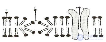 Fig. 4
The selectivity of transfer is provided by a set of pores of a certain radius in the membrane, corresponding to the size of the penetrating particles. This distribution depends on the membrane potential. Thus, the pores in the erythrocyte membrane selective for potassium ions have a comparatively low permeability coefficient equal to 4 pm/s at a membrane potential of 80 mV, which decreases by a factor of 4 as the potential is lowered to 40 mV.
Uncharged molecules of small size (CO2, ethyl alcohol, urea) pass through the membrane quickly, while glucose, amino acids, and fatty acids usually diffuse through membranes slowly. Water diffuses easily through the lipid layer, despite the fact that water molecules are relatively insoluble in fats. This is partly explained by the fact that water molecules are small and uncharged. Specific proteins called membrane transport proteins are responsible for the transfer of various polar molecules, such as sugar, amino acids, nucleotides, and other metabolites, across cell membranes. Each specific protein is designed for the transport of a strictly defined chemical compound. Such carrier proteins are able to bind to a molecule or ion and, without expending energy, that is, passively, transport it across the membrane along the concentration gradient. This process, called facilitated diffusion, is the main mechanism of selective membrane permeability.
Facilitated diffusion occurs with the participation of carrier molecules. For example, valinomycin is a carrier of potassium ions. The valinomycin molecule has the shape of a cuff, inside of which are polar groups, and outside – nonpolar ones (Fig. 5).
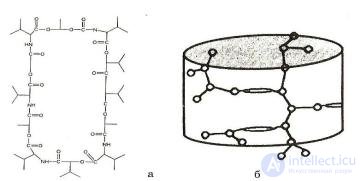 Fig. 5
The valinomycin molecule, owing to its nonpolar group, is able to dissolve in the lipid phase of the membrane and at the same time form a complex with potassium ions entering inside the cuff-molecule. Diffusing within the membrane, the molecules carry potassium across it, and some of them, breaking apart, release potassium ions into the solution on the other side of the membrane. The transfer of potassium across the membrane can occur in both directions. Therefore, if the concentrations of potassium on both sides are the same, the flow of potassium in one direction will be the same as in the other, and as a result no net transfer of potassium across the membrane will occur. But if on one side
the concentration of potassium is greater than on the other (K+ 1 K+ 2), then here the ions will be captured more often
by the carrier molecules than on the other side, and the flow of potassium toward the side of lower K+ will be greater than in the opposite direction (Fig. 6).
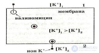
Fig. 6. Thus, facilitated diffusion occurs from places with a higher concentration
of the transported substance to places with a lower concentration. This process explains the transfer across biological membranes of amino acids, sugars, and other biologically important substances.
Facilitated diffusion differs from simple diffusion in the following respects:
1 – transfer of the substance with the participation of a carrier occurs significantly faster;
2 – facilitated diffusion has the property of saturation;
3 – in facilitated diffusion, competition of the transported substances is observed in cases where different substances are transported by the carrier. In this case, the rate of transfer of substances has different values, and the addition of some substances hinders the movement of others. For example, glucose is transported better than fructose, and fructose better than xylose.
4 – the presence of substances that block facilitated diffusion leads to the formation of a stable complex with the carrier molecules; for example, phlorizin suppresses the transport of sugars across the biological membrane
A variant of facilitated diffusion is transport by means of immobile carrier molecules fixed in a certain way across the membrane. In this case, the molecule of the transported substance is passed from one carrier molecule to another, as in a relay.
Active transport of substances, in contrast to facilitated diffusion, occurs against their concentration gradients, that is, substances move from a lower to a higher concentration. In this case, energy is required to transfer protons or inorganic ions across the membrane, the source of which is ATP. According to current concepts, biological membranes contain ion pumps operating on the free energy of ATP hydrolysis – special systems of integral proteins (transport ATPases). The transfer of ions by transport ATPases occurs through the coupling of transfer processes to the energy of cell metabolism.
During the operation of K+-Na+-ATPase, owing to the energy released during the hydrolysis of each ATP molecule, two potassium ions are transported into the cell and at the same time three sodium ions are pumped out of the cell. Thus, a concentration of potassium ions in the cell that is elevated compared with the intercellular medium, and a lowered concentration of sodium, is created. In the course of this process there are seven stages of ion transfer coupled with ATP hydrolysis:
Thus, over a complete cycle, three sodium ions are expelled from the cell, the cytoplasm is enriched with two potassium ions, and one molecule of ATP is hydrolyzed.
Endocytosis and exocytosis. Macromolecules (for example, proteins, polynucleotides, or polysaccharides) enter the cell by means of endocytosis (endo - inside, cyto – cell). Two types of endocytosis are distinguished: phagocytosis (uptake of solid particles) and pinocytosis (uptake of fluids).
During phagocytosis, outgrowths of the cytoplasm surround droplets of fluid with dense particles, for example bacteria, and draw them into the depth of the cytoplasm, where they undergo enzymatic breakdown into fragments that can be absorbed by the cell (Fig. 7).
Owing to their capacity for pinocytosis, cells can take up vesicles of fluid. The process of fluid uptake is similar to phagocytosis. At the point of contact of the droplet with the cell, the plasma membrane forms an invagination in the shape of a small channel, which fills with fluid. It then detaches and enters the cytoplasm, where its membrane walls are digested and its contents are released (Fig. 7). Owing to this process, cells can take up both large molecules and various ions unable to penetrate the membrane because of the extremely small pores for them.
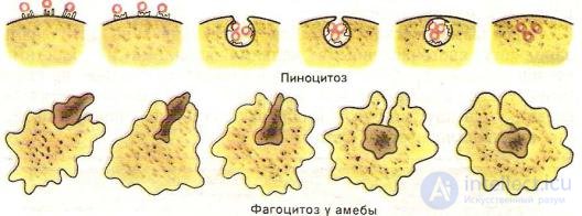
Fig. 7. In the process reverse to endocytosis – exocytosis (exo – outside) – the cell releases the contents
of the vesicle into the external environment. For example, cells producing the hormone insulin package it into intracellular vesicles, which fuse with the plasma membrane and open outward, releasing the insulin in the process.
Osmosis is the predominant movement of water molecules across a semipermeable membrane, which occurs due to a difference (gradient) in concentrations inside the cell and outside it. Osmosis plays a major role in many biological phenomena, for example, it causes hemolysis of erythrocytes in hypotonic solutions.
Filtration occurs through water pores in the membrane under the action of a pressure gradient.
|
The rate of transfer during filtration obeys Poiseuille's law: |
|
||
|
|
dV |
P1 –P2 |
(9) |
|
|
------- = --------- , |
||
|
dV |
dt |
W |
|
|
|
|
|
|
|
where -------- - is the volumetric rate of solution transfer, |
|
||
|
dt |
8 l |
|
|
|
w - hydraulic resistance, |
w =------- , |
|
|
|
|
r4 |
|
|
|
l – pore length, r – pore radius, - viscosity coefficient of the solution, |
P1 –P2 – pressure gradient |
||
|
through the membrane. |
|
|
|
Filtration plays an important role in the transport of small hydrophilic molecules with a radius of less than 4 Å, for example water and urea.
The transformation of toxic substances in the body (metabolism) occurs mainly in two directions: oxidation, often accompanied by hydrolytic cleavage, and reduction. In addition, other reactions also take place: the binding of poisons with proteins, amino acids, glucuronic and sulfuric acids. In most cases, a toxic compound passes through a series of successive transformations, the completion of which are synthetic conjugation reactions (from Latin conjugatio – joining).
The process of transformation of chemical substances in biological media (biotransformation) begins immediately after they enter the body. The initial phase of metabolism is of very great importance, since depending on how it proceeds, the toxicity of the poison may either decrease or increase. Thus, for example, many organophosphorus insecticides undergo oxidation in the body with the formation of more active metabolites: octamethyl is converted into the more toxic phosphoramidoxide; thiophos (parathion) is oxidized to the more active paraoxon. At the same time, the end products of paraoxon (para-nitrophenol and diethyl ester of phosphoric acid) do not possess specific activity.
The study of the metabolism of industrial poisons is important from a practical point of view. Understanding of the mechanisms of biotransformation, of the sequence and rates of transformation
of a given substance can be used for active intervention in these processes, that is, they can be used for prevention, as well as for the purposes of diagnosing intoxication.
Many metabolic reactions are catalyzed by enzymes. Most of them are associated with certain cellular structures (nucleus, mitochondria, lysosomes, and so on). In addition, they are found in the soluble fraction of many tissues and in blood plasma. These same enzymes take part in a number of transformations of ordinary metabolic processes.
Enzymes – substances capable of catalytically influencing the rate of biochemical reactions. They may consist only of protein, or of a protein part and a non-protein part. The non-protein part in a number of enzymes is represented by one or another vitamin.
By the nature of their action, enzymes are divided into exoenzymes, which are released by the cell into the external environment, and endoenzymes, which are firmly bound to the internal structures of the cell and act within it.
Each enzyme possesses strict specificity of action, that is, the ability to affect only certain bonds in complex molecules or only certain substances.
The name of an enzyme consists of a word denoting the substance (substrate) on which it acts and the ending -ase: amylase (acts on starch), protease (acts on proteins), lipase (acts on fats), and so on.
At present more than 1000 enzymes are known. According to the commonality or similarity of their catalytic properties, enzymes are divided into 6 classes:
1.Oxidoreductases - catalyze oxidation-reduction reactions, carrying out the transfer of H and O atoms or electrons from one substance to another, thereby oxidizing the first and reducing the second. This group of enzymes takes part in all processes of biological oxidation.
2.Transferases – catalyze the transfer of a group of atoms (methyl, acyl, phosphate, or amino group) from one substance to another.
3.Hydrolases – accelerate the reaction of splitting complex organic compounds into simpler ones by the addition of water molecules at the site of bond cleavage. This group of enzymes includes amylase (breaks down starch), lipase (breaks down fats), and so on.
4.Lyases – catalyze the non-hydrolytic addition (without the participation of water) to a substrate, or the removal of a group of atoms from it. In this process C–C, C–N, C–O, C–S bonds may be broken.
5.Isomerases – carry out intramolecular rearrangements, that is, they catalyze the conversion of one isomer into another.
6.Ligases (synthetases) – catalyze reactions joining two molecules with the formation of new C–O, C–S, C–N, C–C bonds, using energy.
However, the main significance in the reactions of poison metabolism is attributed to microsomal enzymes. In the cytoplasm of cells there is a very fine network of structures called the endoplasmic network, or endoplasmic reticulum. These structures exist in all animal cells except erythrocytes. The metabolism of poisons occurs in the endoplasmic reticulum of liver cells, since it is there that the highest enzyme activity is observed.
A wide variety of fat-soluble organic compounds of different structure undergo oxidation by microsomal enzymes. These reactions are based on hydroxylation, which is carried out by a series of coupled oxidation-reduction stages. In the oxidation of benzene derivatives, the final reaction is the formation of a sulfoxide. In addition to oxidizing enzymes, the endoplasmic network of the liver also contains enzymes that reduce foreign organic compounds. Aromatic nitro- and azo-compounds and aliphatic halogen-containing compounds undergo reduction. In addition, there are various enzyme systems in the kidneys and lungs that oxidize many primary alcohols to the corresponding aldehydes and further to carboxylic acids. Esters and amides of acids undergo hydrolytic cleavage. Enzymes (esterases, amidases) found in the liver and blood plasma participate in this process:
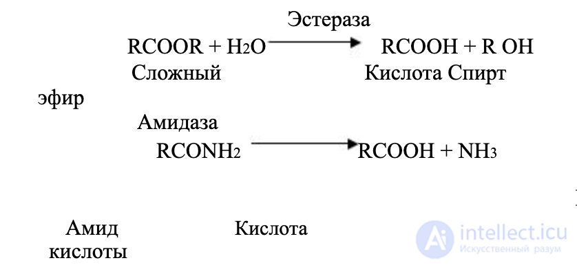
Biotransformation of halogen-containing compounds can also occur through hydrolytic dehalogenation in the liver and kidneys with the formation of chloride or bromide ions and
the corresponding hydrolysis products.

As a result of primary biotransformation reactions, toxic compounds can acquire
chemically active groups (OH, COOH, NH2, SH, and others), which promote further conjugation reactions with readily available compounds: glucuronic acid, sulfate, acetic acid, and certain amino acids. Conjugation leads to the formation of a more polar molecule, which is easily excreted from the body in the urine.
The formation of conjugates is a complex biochemical process, based on the activation of substrates with the participation, in each case, of specific enzymes.
Unlike many organic substances, metals and their compounds, once in the body, can repeatedly change their form. For most of their time in the body, any metals exist in the form of complexes with proteins. Exceptions are the alkali metals and, partly, the alkaline-earth metals. Alkali metals are present in the liquid phase in ionic form, while alkaline-earth metals form unstable, easily hydrolyzed complexes. Metals are also characterized by complex formation with nucleic acids. In addition, metals form complex compounds with proteins and amino acids. It is known that the following combine with amino acids: Hg, Cu, Ni, Pb, Zn, Co, Cd, Mn, Mg, Ca, Ba; predominantly through SH-groups: Hg, Ag, Pb, Cd, Zn, Co; through COOH-groups: Cu, Ni, Zn, Mg, Ca.
The deposition of metals occurs in the form of specific complexes; for example, uranium is deposited in the form of a complex in tissues containing carbonyl and phosphate groups (PO43-), and a lead-protein complex has been found in kidney cells.
Metals with variable valence undergo reduction and oxidation in the body. Thus, pentavalent arsenic is reduced in the body to the more toxic trivalent compound. Hexavalent chromium is reduced to the trivalent form and forms a complex with proteins. Vanadium is reduced from the pentavalent to the trivalent form. Reactions of reduction of manganese and lead in the body are also known.
Examples of biological oxidation are uranium and plutonium. For example, tetravalent plutonium is converted into the hexavalent form.
Substances that are non-electrolytes and have good solubility in fats accumulate in all organs and tissues. In this process, in the first phase of the poison entering the body, blood supply to the organ is the determining factor, which slows the achievement of blood/tissue equilibrium. Subsequently, the main factor influencing the distribution of the poison becomes the sorption capacity of the organ. For fat-soluble substances, the greatest capacity is possessed by adipose tissue and organs rich in lipids (bone marrow, testes, and others). For many lipid-soluble substances, tissue serves as the main depot, retaining the poison both at a higher level and for a longer time than other organs. The duration of retention of poisons in the fat depot is determined by their physicochemical properties. Thus, for example, the natural removal of benzene from adipose tissue after poisoning occurs within 30-48 hours, whereas the insecticide DDT is retained for many months. With the exception of organs and tissues rich in lipids, volatile non-electrolytes are distributed approximately equally in the various tissues of the body.
For metals, no general patterns have been established linking their physicochemical properties with distribution in the body. However, it has been established that metals tend to accumulate in the same tissues where they are normally present as trace elements, or in organs with intensive metabolism (liver, kidneys, endocrine glands). Many heavy metals, upon reaching a cell, become fixed on the cell membrane and thereby disrupt the vital activity of the cell. Metals in the form of soluble and readily dissociating compounds, as well as those prone to forming strong bonds with phosphorus (lead, beryllium, barium, uranium, thorium, and others), accumulate predominantly in bone tissue. Rare-earth metals, in the form of poorly soluble compounds, are selectively retained in the liver, spleen, and bone marrow.
Mercury and cadmium accumulate in the kidneys. For some metals located in groups V – VIII of the periodic system: chromium, vanadium, manganese, cobalt, nickel, arsenic, selenium, a more even distribution in all organs is characteristic.
The routes and mechanisms of elimination of toxic compounds vary. Toxic compounds are excreted through the lungs, kidneys, gastrointestinal tract, and skin. Poisons and their metabolites are often excreted through several channels simultaneously. The elimination of harmful substances from the body proceeds in stages and consists of two or more phases. This is related to the different forms of circulation and deposition of the poison. Compounds that are unchanged or very weakly bound to biological ligands are removed from the body first, then the fraction of the poison found in cells in a more strongly bound form is eliminated, and last of all the poison leaves the body from the tissue depots.
Elimination through the lungs. Most volatile non-electrolytes are excreted from the body mainly unchanged with the exhaled air. Elimination begins immediately after the poison stops entering the body. Initially, the rate of elimination of gases and vapors is determined by their physicochemical properties, and the smaller their solubility coefficient in water, the faster the elimination occurs of the part of the poison that was in the blood and organs. Then the rate of elimination of the harmful substance decreases. This is characteristic of the process of removal of the fraction of the poison deposited in adipose tissue, the quantity of which in a human amounts to about 20% of body weight. For example, during the elimination of chloroform, about 50% of the absorbed compound is exhaled in the first phase, within 8–12 hours, while in the second phase elimination continues for several days.
Volatile metabolites formed during the transformation of the poison can also be excreted through the lungs, since many non-electrolytes form carbon dioxide and water as end products of decomposition.
Elimination through the kidneys. Elimination of harmful substances through the kidneys occurs by two different mechanisms: passive filtration and active transport.
As a result of passive filtration in the renal glomeruli, an ultrafiltrate is formed, which contains the electrolyte at the same concentration as in the plasma. In the renal tubules, non-electrolytes that are highly soluble in lipids can penetrate by passive diffusion in two directions: from the tubules into the blood and from the blood into the tubules. The elimination of volatile non-electrolytes with the urine is insignificant. A quantitative characteristic of the possibility of removing harmful substances through the kidneys is the concentration index K:
|
concentration in urine |
|
|
|
K = ----------------------------- |
(10) |
|
|
concentration in plasma |
|
|
|
The quantitative index for some |
industrial poisons is: methane |
hydrocarbons – about 0.1; chlorinated hydrocarbons (methyl chloride, methylene chloride, chloroform, dichloroethane, trichloroethane, dichloropropane, trichloroethylene) from 0.11 to 1; ketones (acetone, methyl propyl ketone, diethyl ketone) – 1.05 – 1.34; monohydric alcohols (ethyl – 1.3; methyl, propyl, isopropyl alcohols – 1; diethyl ether – 1).
The direction of passive tubular diffusion of weakly ionized organic electrolytes depends on the pH of the urine. If the tubular urine is more alkaline than the plasma, then weak organic acids readily pass into the urine. If the reaction of the urine is more acidic than that of the plasma, then weak organic bases diffuse into it.
In the renal tubules there exist independent systems of active transport for strong organic acids (uric acid) and bases (choline, histamine, and others). Foreign substances of similar structure are excreted from the blood into the urine with the participation of the same carriers. Some industrial poisons containing amino groups in their composition are able to concentrate in the urine.
For example, the concentration of 2-amino-1-naphthol (a metabolite of naphthylamine) in the urine is 200 times higher than in the blood. 2,4-dichlorophenoxyacetic acid is also rapidly excreted in the urine.
Metals present in the form of ions and in a molecularly-dispersed state are rapidly excreted by the kidneys. This applies primarily to the alkali metals excreted with the urine – lithium, rubidium, cesium. Ionizing salts of divalent metals (beryllium, cadmium, copper) are also readily removed with the urine, as are metals present in the body in the form of chelates and metals of groups V – VI, which are part of anions (chromium, vanadium, molybdenum, selenium). Metals retained
predominantly in the liver are excreted with the urine to only a small extent. The elimination of metals through the renal tubules occurs by active transport. Metals distributed evenly throughout the body are removed by two routes: rapidly through the kidneys and more slowly through the gastrointestinal tract. Complex formation promotes the excretion of metals with the urine. This is the basis for the removal of metals from the body using various organic complexing agents.
Elimination through the gastrointestinal tract. The elimination of industrial poisons through the gastrointestinal tract begins already in the mouth, with saliva. Some non-electrolytes and heavy metals, for example mercury, lead, and others, are found in saliva. Swallowing of saliva can return harmful substances to the stomach.
Poisons and their metabolites formed in the liver are transported with bile into the intestine and excreted from the body. Also, absorption of toxic substances directly into the blood can occur from the intestine, with their subsequent excretion from the body in the urine. At the same time, a more complex route is also possible, in which the poison passes from the intestine into the blood and returns again to the liver (enterohepatic circulation).
Volatile non-electrolytes (hydrocarbons, alcohols, ethers, and others) are practically not excreted through the gastrointestinal tract. Chlorinated aromatic and, mainly, polynuclear hydrocarbons (many insecticides) exit through the GI tract. These substances are excreted either unchanged or in the form of their transformation products. Metals are, for the most part, excreted through the gastrointestinal tract. They are retained in the liver and are then excreted with bile through the intestine. Certain metals (lead and manganese) can penetrate directly into the bile.
The form in which a metal is removed through the gastrointestinal tract plays a major role. Metals in the colloidal state are retained in the liver for a long time and are almost completely removed with the feces. These include the light rare-earth metals, gold, silver, and others. The bulk of heavy metals is excreted through the intestine, while their residual amounts are excreted much more slowly with the urine (for example, mercury).
Elimination by other routes. Industrial poisons are also excreted from the body with milk, through the skin, in particular with sweat. Non-electrolytes are excreted with breast milk in animals and humans, for example chlorinated hydrocarbons (insecticides), as well as many metals, for example mercury, selenium, arsenic, and others. Many non-electrolytes are excreted from the body through the skin: ethyl alcohol, acetone, phenol, chlorinated hydrocarbons, and others. Metals such as mercury, copper, and arsenic are excreted with sweat.

Comments