We have come to the moment when we have to move from retelling the basics of neurobiology and the theory of neural networks to the new that the proposed model contains. I advise those who just started reading the cycle to start with the first part.
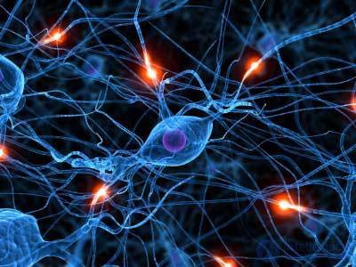
Let's return to the description of the work of real neurons. Signals from some neurons through their axons arrive at the inputs of other neurons. In chemical synapses, a mediator is released, which, depending on the type of synapse, has either an activating or inhibiting effect on the neuron receiving a signal. The sensitivity of the synapse, which can vary, is determined by the contribution of this synapse to the general excitation. If the total impact exceeds a certain threshold, then the membrane depolarizes and the neuron generates a spike. A spike is a single impulse, the duration and amplitude of which does not depend on the synaptic activity that generated it.
The simplest model, inspired by the impulse activity of a neuron, is a model of a threshold adder. At the same time, based on the fact that the spike can be compared with a binary signal, it is assumed that the inputs and output of the adder take values only 0 and 1. If the inputs of such a formal neuron are fed with a pulsed pattern repeated from beat to beat, then the neuron depends on adjusting its weights should either give each response a response, or be silent. This is quite logical - a constant input picture corresponds to a constant result at the output.
If you try to bring the model of the threshold adder to reality, the first thing to do is to assume that the picture of the input activity may not be strictly synchronous. That is, signals at different inputs can be encoded by pulses, each with its own frequency. With this assumption, you can’t just use the instantaneous state of the inputs. It will be necessary to select an indicative time interval and use the activity picture accumulated during this interval. At different frequencies of the input signals, at some intervals, the input pulses will form clusters sufficient to activate the neuron, while at others, they will give discharges, leaving the neuron inactive. Thus, the response of the neuron will acquire its own frequency, which will depend on the frequency of the input signals and the sensitivity of the corresponding synapses.
Such reasoning leads us to a model of a neuron as a linear adder, in which the level of signals at the inputs of a neuron and its response are described not by two levels, but by scalar quantities that correspond to the spike repetition frequencies. The transition to a linear adder allows you to greatly simplify the simulation and partly forget about the original chemical nature of neural activity.
But everything has a beginning. For information to enter the brain, neurons that interact with the outside world are needed. The sensitivity of this interaction is very different from synaptic sensitivity. In synapses, the number of which in a single neuron can be measured in tens of thousands, the mediator is released from the bubbles that have a constant capacity. The minimum quantum of emission of a mediator is a portion of about 7,000 molecules. Sensory neurons work with completely different volumes. Thus, the visual rods are activated literally from two light quanta, the neuron receptors of the olfactory system are able to detect only a few molecules of odorous substance. Such a high susceptibility is achieved through the mechanisms of internal signal amplification.
The interaction of the neuron with the environment occurs due to protein molecules - receptors, which allow external chemical effects to change the state inside the cell. A substance that interacts with a particular type of receptor is called their ligand. For synaptic receptors, ligands are the very neurotransmitters that provide the interaction of neurons.
Neighboring receptors can combine to create dimers (figure below), which in turn, when combined, form receptive clusters.
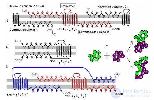 Receptor clustering. A - a single receptor and its interaction with the surrounding receptors. B - monomeric receptive molecule. B - receptive dimer. G is the combination of two monomers into contact (D) and combination (E) dimers. (Radchenko, 2007)
 Receptors are divided into ionotropic and metabotropic. Ionotropic receptors create ion channels that move charged ions across the membrane, changing the membrane potential. When a ligand interacts with an ionotropic receptor, the latter changes the conductivity of the ion channel, opening or closing it. Such receptors are located in the synaptic clefts. Their joint work determines whether or not there is a spike caused by the addition of external signals.
Somewhat different nature of metabotropic receptors. They are located outside the synapses and do not create ion channels. When interacting with their ligand, they trigger the work of intracellular mediators, which provide amplification of the original signal hundreds of thousands of times, which ultimately leads to activation of the neuron. This means that even a small fraction of a substance that is a ligand for the metabotropic receptor can cause a spike in the corresponding neuron. Such a mechanism is used by sensory neurons, it allows them to respond to insignificant third-party effects. But apart from sensory perception, metabotropic receptors are also responsible for a significant proportion of brain activity in general.
A characteristic stimulus for a neuron is a pattern of input signals that coincides with the pattern of sensitivity of its synapses. The closer the input signal is to the characteristic stimulus, the higher the frequency of spikes generated by the neuron. This reaction is called evoked activity. It occurs in all those neurons that have somehow reacted to the current image. But the induced activity is only a small part of the total brain activity. The main activity falls on the so-called background activity (figure below).
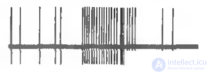 Neuron response to stimulus and background (spontaneous) activity
 Background activity consists of occasional single spikes. Such adhesions are called spontaneous, as they appear regardless of the presence or absence of the induced activity. If the brain is protected from external information, the spontaneous activity on the sensory zones will not weaken, but on the contrary, it will only increase. For an individual neuron, its spontaneous activity looks like a series of random spikes. But for neurons of one zone of the cortex, this activity develops into a general rhythm, which in total creates significant electrical oscillations. These oscillations can be fixed by attaching electrodes to the scalp, which is actually called electroencephalography.
For different zones of the cortex and different states of a person, their own frequencies and levels of such rhythms are characteristic. The rhythm from a separate zone of the cortex is most strongly traced in the area of skin located directly above this zone. Therefore, a set of electrodes uniformly distributed over the surface of the head are used to record brain rhythms. The result looks like a set of graphs, each of which is recorded from its own electrode (figure below).
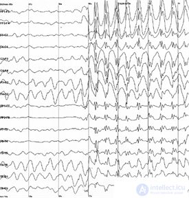 An example of an electroencephalogram. A sharp increase in amplitude corresponds to the onset of an epileptic seizure.
 If the induced activity is a consequence of the work of ionotropic receptors, then spontaneous activity is the result of the triggering of metabotropic receptors. When the impulses of regular or spontaneous activity, spreading along the axon, reach the recipient's neuron, they cause the release of the mediator into the synaptic cleft. This mediator determines the contribution of the synapse to the process of neuron activity caused. Then there is its reverse capture, and the synapse is restored to its original state. But part of the mediator is thrown out of the synaptic cleft and spreads over the space formed by the bodies of neurons and the bodies of the glial cells surrounding them. This phenomenon is called spillover from the English spillover - overflow, overflow. The synapses located nearby from each other form so-called synaptic traps. In these traps, the inhibitory and activating mediators create an interference pattern. This means that each combination of pulses causes an antinode of the mediator in a certain unique place of the membrane. A.N. Radchenko showed that metabotropic receptive clusters located in the places of such antinodes are able to change their properties and subsequently react to the repetition of the same surrounding impulse pattern (Radchenko, 2007).
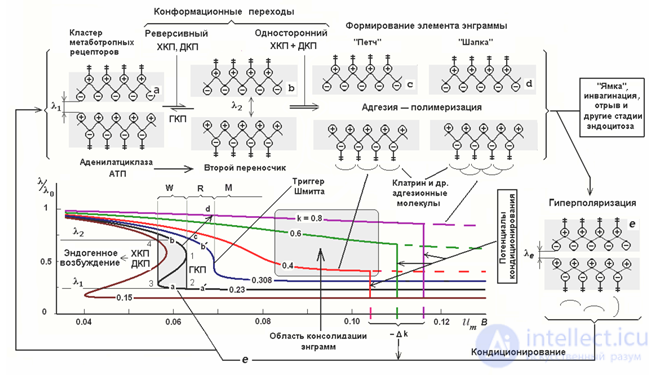 Different states of a metabotropic receptive cluster (Radchenko, 2007)
 Radchenko described several states characteristic of metabotropic receptive clusters and compared these states with different stages of memorization (picture above). The ability of receptors to influence the state of a cell depends on the position of their split ends. The greater the distance between them, the stronger the outer ends protrude on the surface, and the inner ones penetrate the cell environment. Accordingly, a greater distance corresponds to a greater sensitivity of the receptor. When the distance decreases, the ends of the receptors sink into the membrane, and the receptors lose their ability to influence the state of the neuron. The distance can be controlled by the membrane potential of the neuron. Depolarization pushes the end portions out of the membrane to the outside and into the cell, respectively, and hyperpolarization counter-pulls them into the membrane.
Initially metabotropic receptive clusters have hysteresis characteristics. The hysteresis implies that the behavior of the system is determined by its background (chart below). Turning to a certain steady saturated state, the system does not return back after removing the force, but remains in this position. To remove it from this position, it is now necessary to apply the opposite force, which, after passing a certain threshold, will transfer the system to another stable state that has the same “sticking” property.
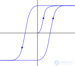 Hysteresis
 Hysteresis gives the receptor properties of a trigger. Reacting to the antinode of the mediator, receptors can “stick” both in the state of ejection from the membrane, and in the state of immersion in it. The first state indicates that the neuron becomes sensitive to a certain picture of environmental activity and may react to its repetition with its own spike, the second, on the contrary, makes it unresponsive to it. But this sticking is short-term, since a certain change in the membrane potential can reset the cluster to its original state.
Radchenko compared this behavior of receptive clusters with short-term (short-term) memory. That is, at each specific moment, our thought determines, on the one hand, a picture of the activity of neurons, and on the other hand, such fixation of traces of this activity, which allows you to quickly restore recent pictures. The fragility of these traces ensures the efficiency of memory. We can quickly return to the thought that was a second or a minute ago, but lose this lightness when hours or days pass.
Under the influence of mediators and a strong shift of the membrane potential, receptors can lose their hysteresis properties and become stable, which permanently fixes the acquired cluster state, which can be compared with long-term memory. At the same time, depending on the position of fixation, the ejected state or recessed, the cluster either reacts to a certain pattern of activity, or vice versa, acquires a stable insensitivity to it. These stable states can be stored for a very long time, one can say forever, although under certain conditions they can also be overwritten.
Further in our model we will proceed from the fact that the brain neurons have three forms of information storage. One form is a change in the sensitivity of synapses, which determines the image characteristic of a neuron. The second form is a short-term trigger fixation of a pattern of environmental activity on metabotropic receptive clusters. And the third is a long-term fixation, due to the transition of receptive clusters to a steady state, parts of briefly fixed pictures.
The sensitivity of synaptic neuron describes the only characteristic image of it (if you do not take into account neurons with several sections of synapses). At the same time, the extra-synaptic surface of a neuron, which contains hundreds of thousands of metabotropic receptive clusters, can store a comparable number of memories indicating in which events this characteristic image appeared.
Note that the impulse activity of neurons consists of two components. One component is a picture of evoked activity, which indicates which neurons have learned their characteristic stimuli. The second component is a picture of conditionally spontaneous activity, which exists relatively independently of the first, and which creates the general background of the brain's work. This background is less accentuated than the induced activity, but more ambitious, since it is distributed throughout the entire space of the cortex.
I’m not afraid to say that it is the background activity that is one of the biggest mysteries of the brain. In the next part we will try to unravel this mystery.
If somewhere is too brief, incomprehensible or vaguely stated, please write in the comments. My eye is already so blurred that it is difficult for me to put myself in the place of the one who perceives this material for the first time. At the same time, if not difficult, indicate the level of your general acquaintance with the topic.

Comments