Lecture
Nerve cells, they are neurons, together with their fibers that transmit signals, form the nervous system. In vertebrates, the main part of neurons is concentrated in the cranial cavity and spinal canal. This is called the central nervous system. Accordingly, the brain and spinal cord are distinguished as its components.
The spinal cord collects signals from most body receptors and transmits them to the brain. Through the structures of the thalamus, they are distributed and projected onto the cortex of the cerebral hemispheres.
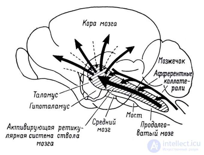
The projection of information on the bark
In addition to the big hemispheres, the cerebellum is also engaged in information processing, which, in fact, is a small independent brain. The cerebellum provides precise motor skills and coordination of all movements.
Sight, hearing and smell provide the brain with a stream of information about the outside world. Each of the components of this stream, passing along its own path, is also projected onto the cortex. The cortex is a layer of gray matter between 1.3 and 4.5 mm thick, constituting the outer surface of the brain. Due to the convolutions formed by the folds, the bark is packed in such a way that it occupies three times less space than in the straightened form. The total area of the cortex of one hemisphere is approximately 7,000 square meters. cm.
As a result, all signals are projected onto the cortex. The projection is carried out by bundles of nerve fibers, which are distributed over limited areas of the cortex. The site on which either external information is projected or information from other parts of the brain forms a zone of the cortex. Depending on what signals come to such a zone, it has its own specialization. Distinguish between the motor zone of the cortex, the sensory zone, the zones of Brock, Wernicke, visual zones, occipital lobe, only about a hundred different zones.
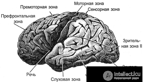
Bark zones
In the vertical direction, the crust can be divided into six layers. These layers do not have clear boundaries and are determined by the predominance of one or another cell type. In different zones of the cortex, these layers can be expressed differently, stronger or weaker. But, in general, we can say that the core is quite universal, and it is assumed that the functioning of its different zones is subject to the same principles.
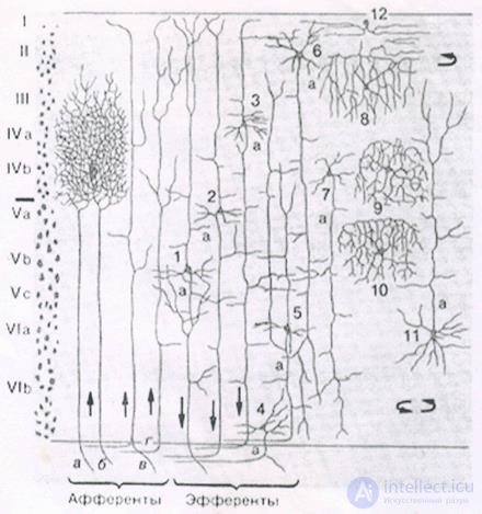
Bark layers
For afferent fibers signals enter the cortex. They fall on the III, IV level of the cortex, where they are distributed in the neurons nearby to the place where the afferent fiber has fallen. Most neurons have axonal connections within their cortex. But some neurons have axons that extend beyond it. For these efferent fibers, signals go either beyond the brain, for example, to the executive organs, or are projected onto other parts of the cortex of their own or of the other hemisphere. Depending on the direction of signal transmission, efferent fibers can be divided into:
If we take the direction perpendicular to the surface of the cortex, it is noticed that the neurons located along this direction react to similar stimuli. Such vertically arranged groups of neurons are called cortical columns.
You can imagine the cerebral cortex as a large canvas, cut into separate zones. The pattern of activity of the neurons in each of the zones encodes certain information. The bundles of nerve fibers, formed by axons that extend beyond their bark, form a system of projection connections. Certain information is projected onto each of the zones. Moreover, several information streams can arrive simultaneously to one zone, which can come from both their own and the opposite hemisphere. Each information flow is similar to a peculiar picture drawn by the activity of the axons of the nerve bundle. The functioning of a separate zone of the cortex is the acquisition of a set of projections, the memorization of information, its processing, the formation of its own picture of activity and the further projection of information resulting from the operation of this zone.
Significant brain volume is white matter. It is formed by axons of neurons, creating those same projection paths. In the figure below, white matter can be seen as a bright filling between the cortex and the internal structures of the brain.
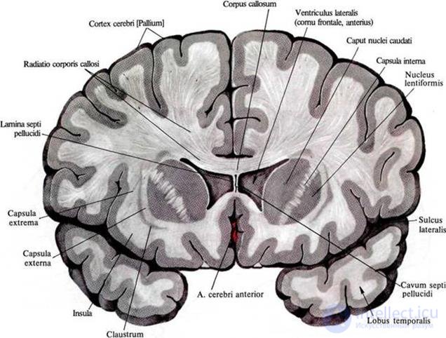
The distribution of white matter in the frontal section of the brain
Using diffuse spectral MRI, we managed to track the direction of individual fibers and build a three-dimensional model of connectivity of the cortex zones (Connectomics project (Connect)).
The structure of relationships is well represented by the figures below (Van J. Wedeen, Douglas L. Rosene, Ruopeng Wang, Guangping Dai, Farzad Mortazavi, Patric Hagmann, Jon H. Kaas, Wen-Yih I. Tseng, 2012).
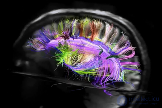
View from the left hemisphere
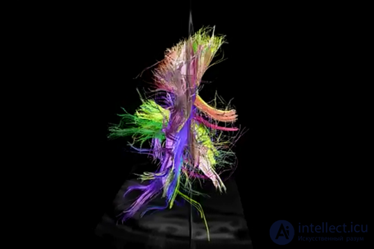
Back view
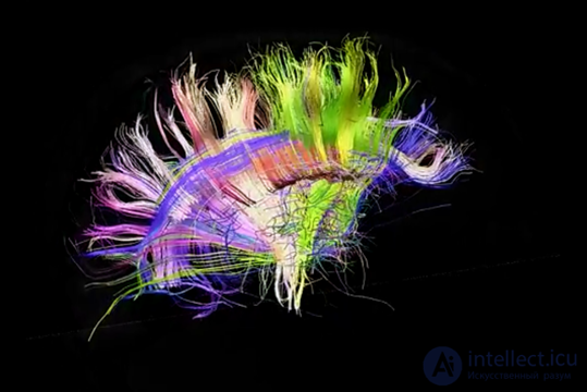
Right view
By the way, on the rear view, the asymmetry of the projection paths of the left and right hemispheres is clearly visible. This asymmetry largely determines the differences in those functions that acquire the hemispheres as they learn.
Comments
To leave a comment
Logic of thinking
Terms: Logic of thinking