
Lecture
Hypogeomagnetic field (HGMF) is an attenuated geomagnetic field - a magnetic field (MF) inside an ordinary or shielded room, or on a planet lacking a magnetic field, which is determined by the superposition of the MF created by the attenuated geomagnetic field, the field from ferromagnetic structural parts of the room, and the field of direct current flowing through busbars, individual structural components, or within them.
Prolonged exposure of a person to HGMF leads to a decline in working capacity and has a negative effect on health. Such fields are a biologically active factor causing a range of changes at the physiological, biochemical, and morphological levels of body function.
The effects of magnetic and electromagnetic fields on biological objects have been studied for quite a long time. And while the mechanisms of action of ionizing radiation have been examined in detail [1, 2], the mechanisms of action of electromagnetic
radiation below the thermal threshold remain unknown to this day.
Of particular interest is the study of the effects of ultra-weak electromagnetic
and magnetic fields whose quantum energy lies below the characteristic
energy of chemical transformation. Data confirming the existence of a biological
effect of such fields are sufficient [3, 4], however the mechanism of the field's action itself
remains unreliably known.
Numerous attempts have been made to explain the physical nature of the biological effects of ultra-weak fields [5–13], however all of them run into the need for experimental confirmation. The absence of clear ideas about experimental design leaves all developments at the level of hypotheses. It is known that the electromagnetic background varies greatly not only in space, but also in time.
One of the main problems faced by experimenters is
the impossibility of standardizing experimental conditions [14]. This factor underlies the poor reproducibility of experiments. Apparently, "zero" magnetic field conditions make it possible to unify the conduct of experiments. In addition,
studies of the "magnetic vacuum" play an important role in investigating the possibility of human adaptation to the conditions of open space.
All living organisms on Earth have been subjected to the geomagnetic field (GMF, ~50 µT), which has constituted the environment of habitation throughout the entire course of evolutionary history. However, the ambient magnetic field (MF) in space geometrically decreases with distance from
Earth. The magnitude of the MF (|B|) at the International Space Station (ISS; near-Earth orbit) is similar to the GMF (~80% of the GMF with limited variation), except that the radial component (Br) has significant variation both in direction and in magnitude. The ambient MF in outer space is extremely lower than the GMF, and is called the hypomagnetic field (HMF); for example, Mars's MF is <5 µT, the Moon's MF is <300 nT, and the interplanetary MF is only a few nanotesla (Figure 1).
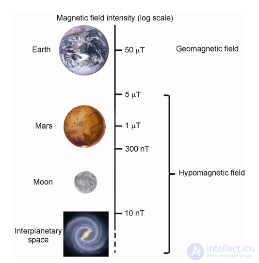
Figure 1. The ambient magnetic field in outer space and the definition of the hypomagnetic field. A weak static magnetic field with |B| <5 µT
is defined as the hypomagnetic field (GMF, HMF). Range of the lunar magnetic field: <300 nT; range of the Martian magnetic field: 300 nT–5 µT. The interplanetary magnetic field is less than 10 nT. Magnetic intensity is presented on a logarithmic scale (modified from a figure in [ ]).
Thus, in addition to microgravity (µ-gravity) and cosmic radiation, astronauts must be exposed to HMF and adapt to these
conditions on board during long-duration and deep-space missions.
Although the biological effects of GMF were assessed in early studies (1960s), research on space GMF has
not progressed rapidly in subsequent decades. There may be several reasons for this. First, given that the strength of the MF in near-Earth orbit is about 80% of the GMF, the effects of microgravity and radiation are more pronounced than those of the GMF in short-term, near-Earth spaceflights.
Much attention has been devoted to ground-based
simulation experiments on microgravity and radiation dosimetry in previous space projects and missions. Second, maintaining a high-quality GMF requires sophisticated instruments and expensive materials, such as a high-precision magnetometer, magnetic shielding metal, and a Helmholtz coil system. In previous studies, the varying conditions of the experimental setups used have often raised
additional problems of reproducibility and comparability [ ]. Third, the effects of GMF are weak or reversible in many
cases, although some biological responses have been found
to be notable in recent years. It has been suggested that gravity may mask the effects of GMF in ground-based HMF simulation experiments. Unfortunately, reports on GMF testing aboard the ISS remain very limited to date. Overlooked HMF effects may indeed pose a substantial risk to the health of astronauts during long journeys in open space.
Substantial results on the effects of GMF have been published in the last decade. A number of experiments have made it evident that GMF significantly disrupts the functional state of organisms [2,3]. It has been shown that exposure to GMF alters embryonic development, cell proliferation, the state of oxidative stress, and immune system function [2,3].
In animals/humans exposed to GMF, impairments of learning and memory, and declines in cognitive ability and working capacity have been observed. We recently performed a transcriptomic profile analysis of human neuroblastoma cells exposed to GMF and found that GMF exposure significantly affects the transcription of genes related to macromolecule transport, metabolic processes, and mRNA processing, as well as downstream pathways involved in cytoskeletal organization, regulation of chromatin condensation, transcription, and brain function [ ]. Given reports of the adverse effects of HMF on many aspects of living systems, especially brain function, astronauts exposed to GMF are consequently subject to potential health risks during interplanetary flight navigation. Thus, it is necessary to elucidate the mechanism
of the GMF effect and to develop effective strategies to counteract GMF for the life support and health care of astronauts in space.
In fact, space μ-gravity, radiation, and HMF act on multiple aspects of the organism, including bone metastasis,
the brain and cognition, the immune system, cell growth and embryonic development, the cytoskeleton, the stability of genetic material (e.g., epigenetic changes), and responses to oxidative stress (Table S1 in the Supplementary Information), which implies the existence of interactions among the three factors.
Current experiments have considered the combined effects of radiation with gravity, while the interaction of HMF with radiation or gravity on the organism has not received due attention. However, in ground-based apparatus studies, the effects of μ-gravity
and GMF are in fact often confounded with GMF and normal gravity, respectively. For example, the potential effect of GMF is largely overlooked in observations of gravity effects using conventional μ-gravity simulation approaches (e.g., the 3D clinostat). Moreover, we observed enhanced gravitropism in germinating soybean seeds by eliminating interference from GMF under a ground-based GMF simulation system [ ]. Therefore, it would also be advisable to clarify the influence of GMF when investigating the biological effect of μ-gravity in ground-based
experiments, for example, by installing a 2D or 3D clinostat inside a magnetically shielded chamber (GMF environment), and vice versa (Figure 2). Thus, the magneto-gravitational effect suggests both the necessity and the opportunity to initiate research on
the combined effects of μ-gravity, radiation, and HMF in further scientific projects, in addition to the practical need to anticipate real risk in space.
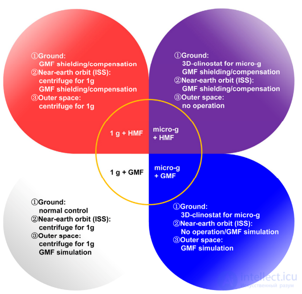
Figure 2. Experimental concept for elucidating the biological effects of μ -gravity and GMF in various experimental environments.
Biological responses to the three factors should be carefully distinguished using combined experimental setups that
extend their capabilities beyond simply simulating a "true" space environment. Designing a feasible, practical, and reliable experimental setup that can accurately provide complex combinations of ionizing radiation, μ -gravity, and/or GMF is crucial for improving progress in space life science. Close collaboration between experienced biologists and engineers will be highly valuable and necessary.
To understand the biological action of an attenuated geomagnetic field, researchers apply two fundamentally different approaches. The first is based on the principle of active compensation of the geomagnetic field, for example using three pairs of mutually perpendicular coils (Helmholtz coils), and consists in creating a magnetic field equal in magnitude but opposite in direction to the geomagnetic field.
The disadvantages of this approach are as follows: first, the region of practically uniform
compensated magnetic field is extremely small, amounting to ∼20% of the volume
bounded by the coils, which seriously complicates placing a laboratory animal there; second, the biological object itself is completely exposed to all manner of interference from man-made alternating electromagnetic fields at 50–100 and 400 Hz,
as well as high-frequency interference.
The second approach is shielding an enclosed volume with a material of high magnetic permeability. In this case, man-made interference is not a concern; however, when amorphous soft magnetic materials are used as shields, biological objects must be placed along the axis of shielding cylinders or at the center of shielding spheres, dictated by the particular way magnetic field
lines pass through such materials.
Both of these approaches were used in our studies. When studying the effect of an attenuated geomagnetic field at the level of the whole organism, a laboratory animal - an SHR-line rat - was placed in the compensation setup. It was found that under compensated-field conditions the animal exhibited sharp fluctuations in blood pressure (BP) and heart rate (HR), but it was not possible to systematize these effects owing to the influence of individual characteristics of the laboratory animals on the degree and nature of the manifestation of the magnetobiological effect. The difficulties in interpreting results at the level of the whole organism served as the motivation for conducting experiments at a biological level that is simpler in organization.
Two cell lines were chosen as models: the HeLa epithelioid carcinoma line and primary human VH-10 fibroblasts. It was assumed that cells growing under normal geomagnetic field conditions would perceive its shielding as a stress factor and possibly exhibit a classic cellular response to damage. By now the mechanism of the global cellular response
has been well studied [15]: most of the genes whose products are involved in it have been cloned and sequenced. The main protein involved in the processes of maintaining cellular stability is the anti-oncogene P-53, the appearance of which in phosphorylated form can serve as a marker for the triggering of the global cellular response
to damage [16–18].
It is known that any cellular change requires energy resources. The source of ATP in the cell is the mitochondria, which in normally proliferating
cells of a culture form a network that performs an integrating function in the cell’s «energy system.» Study [19] showed that the structure of the mitochondrial
network is extremely plastic and capable of reorganization under the action of damaging agents. Thus, for a preliminary analysis of the cellular response to a hypogeomagnetic field, we chose to investigate the P-53 status and the mitochondrial
network of the cells under study.
Compensation of the geomagnetic field. Compensation of the geomagnetic field was carried out using three pairs of mutually perpendicular Helmholtz coils. To
control the currents in the coil windings of the setup, a three-channel current amplifier was designed and built, allowing separate regulation of each of the three components
of the magnetic field induction vector.
Shielding of the geomagnetic field. To study the effect of extremely weak
magnetic fields on biological objects at the cellular and subcellular levels, a
shielding chamber was created, representing a cylinder (D = 26 cm, L = 84 cm),
covered with dozens of layers of shielding material made from an alloy based on the amorphous soft magnetic material AMAG-172. The total number of layers is 40. All layers are «wound» in one direction and grouped into 4 independent packets
of 10 layers each, with 3 mm air gaps between the packets. At one end of the cylinder there is a fixed plug, at the other — a removable lid. The plug and lid have a shielding coating identical to that of the cylinder. The design of the removable lid avoids the appearance of «magnetic holes» in the shield.
Inside the chamber there is a stand made of non-magnetic material, allowing
biological objects to be placed at the center of the shielding chamber. The multilayer
structure of the magnetic shield made it possible to achieve a shielding factor of 250 for the constant component of the external magnetic field, attenuating the magnetic
field of the Earth (48 µT) down to 0.192 µT at the center of the closed chamber. Taking
into account the frequency characteristics of the soft magnetic material AMAG-172 declared by the manufacturer, it should be noted that there is also a high degree of shielding from any
high-frequency interference and pickup.
Instruments for measurements and data processing. Measurements of the magnetic fields
were carried out with a single-axis FLUXMASTER magnetometer (1 nT to 200 µT)
manufactured in Germany, as well as a three-axis HB0302.1A magnetometer (Russia) with a resolution of 0.1 µT, equipped with specially developed
software for a PC (personal computer) for monitoring both the
individual components and the total vector of the magnetic field in real time. To record the variable components of the electromagnetic field in the working
volumes, NV-0303 magnetic field induction transducers (sensors)
and NV-0303.1 are used, with a working frequency range (flat portion of the frequency response) of, respectively,
5–10000 Hz and 1–1000 kHz. To acquire data from these sensors (digitization and
input of the signal into the computer), a PCS-500 PC oscilloscope from Welleman
(Belgium) is used. Analysis of the frequency spectrum was carried out using the software package
PC-Lab 2000SE.
Studies of the effect of the magnetic field on the homeostasis of the cardiovascular system of the biological subject (laboratory rat): 1) blood pressure (BP) and its variability;
2) heart rate (HR) and its variability — depending on the initial state (BP level, type
of anesthesia, state of the antioxidant system) of this subject — were carried out using
an original BP and HR measurement system built on the basis of a sensor
for direct measurement of arterial blood pressure by Baxter, and the original computer program KardioPlus, which allows evaluation of the heart contraction rate,
as well as changes in the influence of various regulatory systems on rhythm variability.
Microscopy and image analysis. Analysis of cells fixed on glass
slides was performed using an LSM-5 Pascal laser scanning confocal microscope (C. Zeiss, Germany), equipped with a 63/1.4 objective and an argon laser (458/488 nm).
Statistical processing of the results. Statistical processing and
presentation of graphs were carried out using the Excel software package (Microsoft).
Model systems. For the experiments under compensation conditions, both the widely known Wistar-line rats (living systems) and the standard model object for
studying the pharmacological action of antihypertensive drugs — hypertensive SHR-line rats — were used. WKY-line rats, close to the experimental line, were used as controls.
The animal was placed on a non-magnetic table at the geometric center of the setup,
where the field is most uniform. The rat was anesthetized with an intraperitoneal injection of sodium oxybutyrate solution. The HR and BP sensor was inserted into the animal’s femoral artery,
and the signal from it was fed to an amplifier and, through an analog-to-digital converter
(RPG-VCh), to a personal computer, where it was analyzed.
Cells were grown in plastic flasks in Petri dishes (Nunclon, USA)
and on glass slides placed in a Petri dish, in F-10 or DMEM medium (Biolot, Russia, or Sigma, USA) with the addition of 10% fetal bovine
serum (Sigma) and antibiotics (100 U/mL penicillin, 100 µg/mL strepto101
mycin) at 37◦C in an atmosphere containing 5% CO2. HeLa cells — an epithelioid
human carcinoma obtained from the cell culture collection of the Institute of Cytology, Russian Academy of Sciences, were used. The VH-10 cell strain — diploid foreskin fibroblasts from an 11-year-old boy, used in the study as cells from a healthy donor [20, 21].
The cells were kindly provided by Professor A. Kolman (A. Kolman, Stockholm
University, Sweden).
P-53 detection. Immunofluorescence analysis of P-53 was performed on fixed cells. Cells grown on coverslips to a subconfluent state
were fixed with a 4% formaldehyde solution in PBS (phosphate buffer) on
ice for 10 min. After intensive washing with PBS, the cells were permeabilized
in a 0.5% solution of Triton X-100 (Sigma) in PBS for 5 min, washed with PBS, and
placed for 30 min in a 1% solution of bovine serum albumin (BSA, Sigma)
in PBS. To visualize the wild type and most mutant forms of the P-53 protein by indirect immunofluorescence, the cells were first incubated for 60 min with
commercial polyclonal rabbit antibodies against human P-53 protein
(1:50, Santa Cruz, USA), then — for 30 min with goat anti-rabbit gamma globulin antibodies conjugated with fluorescein isothiocyanate (FITC, Sigma), at a dilution of 1:300. Between antibody incubations, the slides were washed for 30 min in a 0.1%
solution of Tween 20 (Sigma) in PBS. After staining, the preparations were mounted in a solution
of propyl gallate in 90% glycerol, which prevents fluorescence fading.
Study of the mitochondrial network. Staining of mitochondria was performed on living cells. HeLa and VH-10 cell lines were grown on coverslips in Petri dishes. Mitochondria were stained with the commercial dyes
MitoTrackerr Orange CM-H2TMRos and MitoTrackerr Green FM (Molecular Probes, USA),
which fluoresced only upon entering the mitochondrion. The dye was incubated with the cell culture at concentrations of Orange — 100 nM, Green — 50 nM, for
40 min in an incubator, then washed with fresh culture medium. After staining, the preparations were mounted in a solution of propyl gallate in 90% glycerol, which prevents fluorescence fading. Cells were placed in the shielding chamber for 0.5, 1,
and 3 h, while control cell cultures remained outside the chamber; the exposure was carried out in a thermostat at 37◦C with a pre-warmed chamber. At the specified time intervals, experimental and control cells were stained while alive with
MitoTrackerr Orange CM-H2TMRos and MitoTrackerr Green. The number of cells
with a reorganizing mitochondrial network was visually counted in 10 fields of view
per preparation, at a comparably uniform seeding density. Tables of
cell counts versus exposure time in the chamber were compiled.
Treatment with H2O2. Cells were treated with H2O2 at a concentration of 500 µM for 1 h before staining under standard culture conditions.
Review of research results in the field of vision, memory, and cognition in the fruit fly Drosophila melanogaster over recent years. Cognitive aspects of perception, learning, memory, and decision-making are distinguished within the framework of minimal intelligence at a lower level, as opposed to a high level of intelligence.
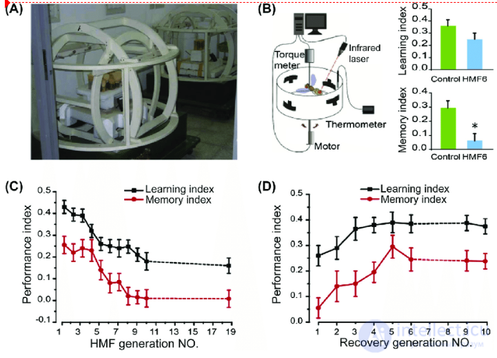
Cognitive deficits in wild-type fruit flies raised in a hypomagnetic field (HMF). (A) Helmholtz coils for compensating the geomagnetic field (GMF), with flies raised in the central part. (B) Visual learning and memory were tested using a flight simulator. The memory index of the sixth generation of HMF flies (HMF6) was significantly reduced (P <0.01). (C) The learning and memory scores of HMF flies progressively declined as the number of generations in the HMF increased. (D) When the 10th-generation HMF flies were returned to a normal GMF, learning and memory ability gradually recovered. Adapted and modified with permission from Zhang, B., Lu, H., Xi, W., Zhou, X., Xu, S., Zhang, K., Jiang, J., Li, Y., Guo, A., 2004. Multiple-generation exposure to a spaceflight hypomagnetic field induces amnesia in Drosophila melanogaster. Neurosci. Lett.
It has previously been shown that extremely weak magnetic fields presumably have pleiotropic mechanisms of action on microscopic fungi. The aim of this work was to investigate the combined effect of shielded extremely weak magnetic fields (B ≈ 100 nT) and antifungal drugs, differing in their cellular targets of action, on colonies of microscopic fungi. The following were selected: (i) polyene antibiotics — amphotericin B (AMP-B) and nystatin, which form complexes with ergosterol and disrupt the plasma membrane of fungal cells, leading to an increase in its permeability, leakage of ions from the cytoplasm, and death of the fungal cell; (ii) azoles – clotrimazole, itraconazole, fluconazole, which inhibit the fungal enzyme C14-α-demethylase of the cytochrome P450 system, responsible for the conversion of lanosterol to ergosterol. This leads to depletion of ergosterol in the fungal cell membrane and its death.
The study used the fungi Ulocladium consortiale, Aspergillus sp., Trichoderma sp., Mucor sp., Penicillium sp. Two alternative methods for testing fungicides were used: first, the standard Kirby-Bauer disk-diffusion method with assessment of the fungal growth inhibition zone size, in which Petri dishes were inoculated with fungal spores as a continuous lawn; and second, growth rate was assessed by the increase in colony diameter upon inoculation at the center of a Petri dish, on which two disks were placed along an axis, as in the first experimental variant. In both inoculation variants, the fungi were cultivated under hypomagnetic field conditions (in a shielding chamber at B ≈ 100 nT) and in the Earth's field under identical illumination and temperature conditions in the control and experimental groups.
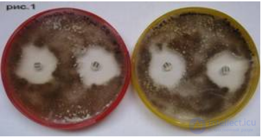
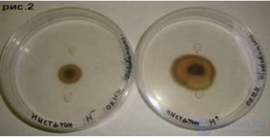
The results of the diffusion method showed that the sensitivity of the fungi to antibiotics, both in the control and in the experiment, decreases in the series nystatin > itraconazole > AMB > fluconazole > clotrimazole, with no statistically significant differences found in the diameters of the inhibition zones between hypomagnetic conditions and the geomagnetic field (see, for example, Fig. 1). However, for the center-inoculation method, statistically significant differences were found for AMB and nystatin between the mean colony growth rate values along the line between the antibiotic disks in the control and experimental samples (p=0.01474<0.05). For example, for disks with AMB, the specific growth rate of U. consortiale was μEMF = 3.345± 0.108 mm/day, μHMF = 2.293± 0.308 mm/day (see Fig. 2). Thus, the experimental results demonstrated that deep shielding of the Earth's magnetic field affects the colony growth rate (but not its final size) in the presence of polyene-type antibiotics (AMB and nystatin). It can be assumed that the target of the magnetic field may be the hydrogen bonds formed when macrolide antibiotics bind to ergosterol in the cell membrane of micromycetes, leading to a synergistic effect.
|
|
|
|
Fig. 1. Identical growth inhibition zones of micromycetes by nystatin under cultivation conditions: left - hypomagnetic field, right - Earth's magnetic field. |
Fig. 2. Differences in the growth rate of micromycete colonies in the presence of nystatin disks. left - hypomagnetic field, right - Earth's magnetic field. |
Laboratory animals. The experiments performed showed that the dynamics of the recorded parameters in hypertensive and normotensive rats under the influence of the compensating field are directed in opposite directions. In normotensive animals, an increase in blood pressure was observed (from 140
to 163 mmHg), followed by a slow decrease to the baseline level. In parallel (and possibly preemptively), an increase in heart rate was observed. And although blood pressure returned to baseline after 100–110 min
, the heart rate remained elevated.
Under the same conditions, in the hypertensive rat (SHR line), blood pressure under the influence of the compensating field decreased monotonically (from 165 to
137 mmHg). At the same time, an increase in heart rate was observed. The moment the compensating field was switched off was accompanied by the development of arrhythmia and a sharp
decrease in blood pressure. Over the following 100–120 min, the recorded parameters returned to baseline.
However, these deviations were irregular and poorly reproducible across different experimental series. This is probably because the response of the whole organism is complex, and its various systems mutually compensate for the stressor effect in order to preserve overall homeostasis. Thus, it is quite difficult to identify the target of the field's action at the organismal level. This prompted the search for
a more simply organized model, an example of which is cell culture.
Cell cultures. Cell cultures are now the main model for studying the effects of various factors on biological objects.
To date, a fairly detailed understanding has been formulated of the activation and interaction of various cell signaling pathways in response to stressor
conditions. Cellular responses to DNA damage have been studied most fully.
Most DNA damage does not result solely from replication errors. Numerous lesions arise at any time during the cell cycle under the action of both exogenous and endogenous factors. Thus, ultraviolet radiation causes the formation of pyrimidine dimers, 6,4-photoproducts, adducts, breaks
and other DNA damage. Under the action of chemical agents, various
kinds of nucleotide modifications occur, interstrand crosslinks arise, and conformational
defects [22].
The effects of ionizing X-ray radiation have been studied in the greatest detail [1, 2]; as a result of this exposure, double-strand breaks arise in the DNA molecule
[23, 24]. These can lead to cell death or stable chromosomal rearrangements [25–27], which is the main cause of the lethal and mutagenic effects
of ionizing radiation. The appearance of double-strand breaks in DNA triggers a cascade
of intracellular reactions mediated by various signaling pathways and leads to the development of a global cellular response to damage, including the activation of specific checkpoints arising in response to DNA damage. The consequences of this include possible cell cycle arrest, enhancement of repair
processes, and changes in chromatin conformation [1, 25–27].
It is known that any cellular change requires energy resources. The source of ATP in the cell is mitochondria, which in normally proliferating
culture cells form a network that performs an integrating function in the cell's «energy system». It has been shown [19] that the structure of the mitochondrial network is extremely plastic and capable of reorganization in response to damaging agents. As a model, we studied two cell types — normal and
tumor cells, since the response of these cell types to damage may differ.
As a marker of the cellular response to stressor effects, the protein
p53 was chosen, since it is the main protein involved in maintaining
cellular stability, the appearance of which in phosphorylated form can serve as a marker of the onset of the global cellular response to DNA damage [16–18], and
the state of the mitochondrial network of the studied cells.
The study of the effect of hypogeomagnetic conditions on HeLa and VH-10 cell cultures
was carried out in two stages. The first step was to determine the presence of p53 protein in detectable amounts. One hour after exposure of the cells in the shielded
geomagnetic conditions, both in HeLa and VH-10 cells, a bright specific green fluorescence is detected in the nuclei, corresponding to the appearance of detectable
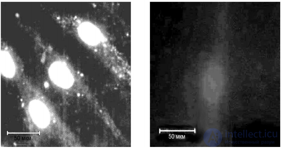
Fig. 1. Detection of p53 protein in VH-10 culture cells:
A - p53 fluorescence in the nuclei of cells exposed for 1 h under shielding conditions; B -
absence of fluorescence in control cells
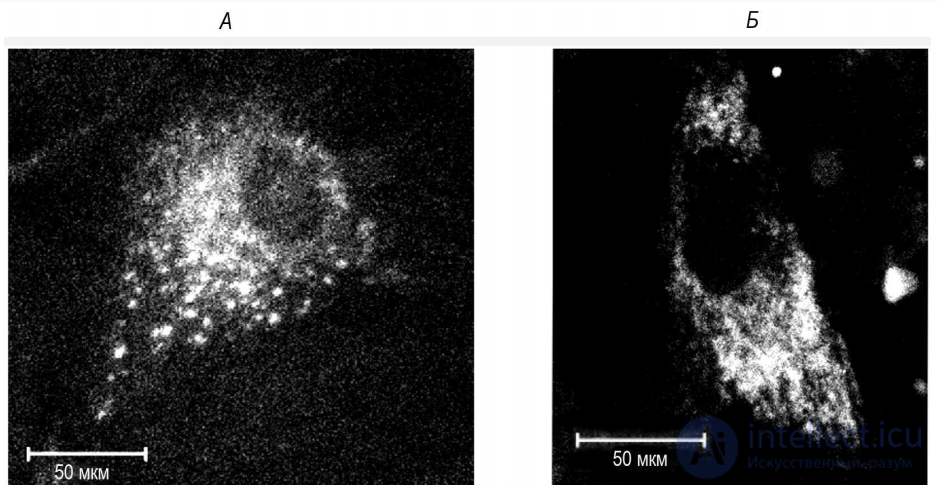
Fig. 2. Detection of the mitochondrial network with the intercalating dye MitoTracker
Green FM
VH-10 cell culture. A - control: ordered, regular mitochondrial network;
B - state of the mitochondrial network after three-hour exposure under shielding conditions
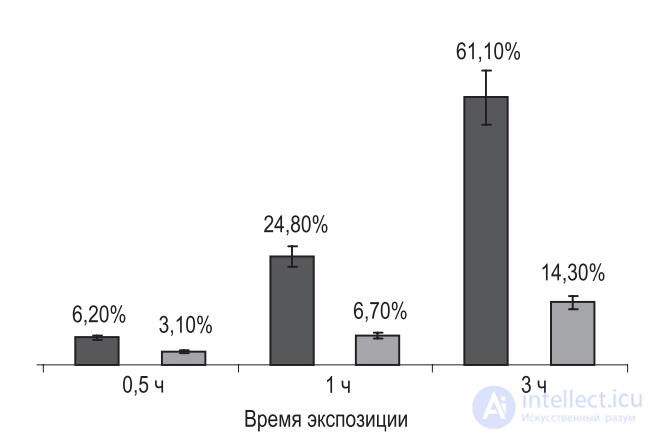
Fig. 3. Average number of cells in culture (in
percent) with a reorganized mitochondrial network after 0.5, 1, and 3 h of exposure under hypomagnetic
conditions
Dark bars — experiment, light bars — control
data by the p53 protein quantification method (Fig. 1). This indicates the stabilization
of the p53 protein in the cell nucleus and shows that p53 and the signaling pathways it mediates
are actively involved in cellular adaptation to conditions of reduced geomagnetic field.
The observed effect was the same in both cell lines, but in further work we
will continue working with primary VH-10 fibroblasts, since these cells are larger and
the results are more clearly visible.
The next step was to determine the state of the mitochondrial network in the control
and after various exposures under shielded geomagnetic field conditions. In
all cells exposed for 1 and 3 h under shielding conditions, a
reorganization of the mitochondrial network, which is normally ordered and located
predominantly around the nucleus (see Fig. 1; 2), was observed. Already one hour after the cells were placed
under the altered conditions, the mitochondrial network broke down, forming both isolated
clusters and large irregular conglomerates in the cell cytoplasm (Fig. 1, A;
2, B). The number of cells with such a reorganized network increased with increasing
exposure time (Fig. 3). It should be noted that cells with an irregular mitochondrial network were also occasionally found
in the control group, but their number was small.
1. Under conditions of a shielded geomagnetic field in HeLa and VH-10 cell lines,
it has been shown that the p53 protein and the signaling pathways it mediates are actively involved in
the cell's adaptation to hypogeomagnetic field conditions.
2. The effect found, based on the parameters studied, proved to be similar to the cellular response to DNA damage.
∗ ∗ ∗
The authors express their deep gratitude for the cooperation and assistance of the staff of the Laboratory of Experimental Physiology and Pharmacology of the «Almazov Federal
Center for Heart, Blood, and Endocrinology» and to colleagues from the Institute
of Structural Materials «Prometey».
∗This work was supported by the Russian Foundation for Basic Research (RFBR) (grant No. 09-04-01208), the Ministry of Education and
Science of Russia (grant No. 2.1.1/485), and the PRAN program (Gene Pools and Genetic Diversity).
c M. L. Kuranova, A. E. Pavlov, I. M. Spivak, S. V. Surma, B. F. Shchegolev, P. A. Kuznetsov,
V. E. Stefanov, 2010

Comments