
Lecture
An engram is a unit of cognitive information imprinted in a physical substance, theoretically the means by which memories are stored as biophysical or biochemical changes in the brain or other biological tissue in response to external stimuli.
Demonstrating the existence, as well as the precise mechanism and location, of neurologically defined engrams has been the focus of ongoing research for many decades.
Engram (Greek έν — remaining in some state or action + Greek γράμμα — everything written, a record), engram , engram — a term introduced into circulation by the German zoologist and biologist Richard Semon at the beginning of the 20th century. In the theory of mnemism, an engram is a physical habit or memory trace on the protoplasm of an organism, left by the repeated action of a stimulus.
Literally, from the Greek, the word "engram" means "inner record". The ancient Greeks used this word to denote wax tablets for recording the meaning of various signs.
The term "engram" was introduced by the memory researcher Richard Semon in reference to the physical substrate of memory in the brain. One of the first undertakings aimed at localizing a memory in the brain was made by Karl S. Lashley, who removed portions of the brain in rodents. In Lashley's experiments, rats were trained to run a maze, and then tissue was removed from their cerebral cortex. Increasing the amount of tissue removed increased the degradation of memory, but, more notably, where the tissue was removed made no difference. Thus, his search proved unsuccessful, and his conclusion that memory is distributed diffusely throughout the brain became widely influential. Today, however, we understand that memory is not entirely but only to a considerable extent distributed throughout the brain; this, together with its dynamic nature, makes it difficult to identify engrams using traditional scientific methods.
Later, Richard F. Thompson searched for the engram in the cerebellum rather than in the cerebral cortex. He used classical conditioning of the eyelid response in rabbits in his search for the engram. He puffed air onto the cornea of the eye and paired it with a tone. After a series of trials involving this sound, the rabbits learned to blink when they heard the sound, even without the puff. One of the areas studied by Thompson's group was the lateral interpositus nucleus (LIP). When it was chemically deactivated, the conditioned response disappeared; when it was reactivated, they responded again, demonstrating that the LIP is a key element of the engram for this response. This approach, targeting the cerebellum, although successful, investigates only basic, automatic responses that virtually all animals possess. However, the engrams of particular types of memory are found in the subsystems mediating that learning process, and as such only engrams of simple conditioning are associated with the LIP, but not, for example, engrams of semantic memory.
Since the beginning of the last century, researchers have tried to explain the neurobiological foundations of memory. Everything we do, everything we think and feel, is based on neural mechanisms, and this necessarily includes memory. But the engram — the presumed neural trace of a memory — is notoriously difficult to pin down.
In large part this is because memory is so pervasive that it is hard to give examples of brain functions that are not related to memory. This means that extensive brain networks will somehow be involved in memory. What we do know (rather unhelpfully) is that it is not located in one particular place or even in several places. The hippocampus is known as the seat of memory, but that is only a small part of the story; the Wikipedia page for the neuroanatomy of memory lists the hippocampus, cerebellum, amygdala, basal ganglia, frontal lobe, temporal, parietal and occipital lobes as parts of the brain associated with memory function. Well, that is nearly every part of the brain involved in cognition… at all.
The first hint that the key to the engram might be neural networks rather than chunks of brain tissue was an experiment conducted by Lashley in 1929. He placed rats in a series of mazes of varying complexity and gave them plenty of time to learn the layout (rats are very good at this). Then he made lesions in their brains to see what would happen. He found that it did not matter where he made the lesion, the rat would still retain some.Some memory remained of how to navigate the maze. What did matter was how large the lesion was — the higher the percentage of cortical brain tissue he cut out, the more errors the rat made (an error was counted as a wrong turn). This study was rather revolutionary for the then still young science of neuropsychology because of the implications for how the brain is organized. There are few centres for various cognitive abilities; brain functions are often spread across extensive brain networks.
(In fact, if you repeated Lashley's experiment more precisely, you would find that there are subtle differences and variations in severity when you cut out different parts of the cortex. Lashley's mistake was that his lesions were so large that each one damaged several cortical areas involved in memory and spatial navigation. In any case, if you disrupt one of the forms of memory involved in the maze task — say, navigation by smell — this can be compensated for by another method, such as vision.)
So, memory is mediated by networks of neurons. How did this come about? How do you store a memory? We do not fully know, but there are some explanations that may enlighten us. One of them is the idea of long-term potentiation, the modification of synaptic strength. Synapses are the chemical junctions between neurons. Essentially, they are the gaps between brain cells that work by transmitting neurotransmitters (chemicals such as serotonin) released by one neuron to the next neuron, and then that neuron generates an electrical impulse. When the electrical impulse reaches the other end of the neuron, it releases neurotransmitter into the next synapse, and so on.
In 1966, a Norwegian researcher named Lømo discovered that if you repeatedly stimulate a nerve pathway with pulses of electricity at high frequency, the synapses in that pathway become more efficient — less effort is required to transmit the signal the next time, because the resulting electrical potential in the second neuron is about 50% greater. He did this on the neurons of the perforant path, which is associated with memory and projects to the hippocampus. The effect lasted up to 16 weeks in unanaesthetized rabbits used as the subjects of his experiment.
External events are represented in the brain as spatiotemporal patterns of neural activation. This is the fundamental premise of neuropsychology; everything we experience is associated with these neural patterns. This means that all learning and memory must be represented by synaptic changes, otherwise nothing in our experience would change, and that would mean we had remembered nothing. Lømo's discovery was the first empirical confirmation of this, and since then we have learned more about how synapses can change their strength.
And here comes the tricky part, so pay attention. In neuroscience there is a general rule that neurons that fire together wire together — this is the basis of Hebbian learning. As the intensity of the input increases, so does both the probability that long-term potentiation will occur and the magnitude of the LTP. There is a threshold of intensity that must be reached in order for synaptic changes to occur, and above this threshold higher intensities lead to greater LTP. What is the significance of this? It means that a minimum number of synapses must act jointly — fire together — in order for LTP to arise. This is how certain neural circuits begin to "form" a memory: when there is the right type of input, for example, the neural processing of the sight of something that posed a threat to you in the past, that particular circuit of neurons begins to activate more readily than it would if you did not have such a memory. In effect, your brain is replaying the information. That is what a memory trace is; it is the mysterious engram.
Stimuli or irritants leave distinct traces (engrams) on the protoplasm of an animal or plant. When the action of this stimulus is regularly repeated, this creates a habit that remains in the protoplasm after the action of the stimulus has ceased.
A hypothetical memory trace. By content, two kinds of engrams are distinguished: images (a representation of the static structure of an object) and action models (programmes).
By hierarchical levels of complexity, a distinction is made between primary engrams, associations of engrams (two interconnected engrams) and associative networks of engrams.
Neurobiology recognizes the existence of many types of memory, and their physical location in the brain is likely to depend on the corresponding system mediating the encoding of that memory. Parts of the brain such as the cerebellum, the striatum, the cerebral cortex, the hippocampus and the amygdala are believed to play an important role in memory. For example, the hippocampus is believed to be involved in spatial and declarative memory, as well as in consolidating short-term memory into long-term memory.
Research has shown that declarative memories move between the limbic system, deep within the brain, and outer, cortical areas. They differ from the mechanisms of the more primitive cerebellum, which dominates the blink response and directly receives auditory input. There is no need to "turn" to other brain structures for help in forming certain memories of simple associations.
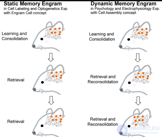
A study at the Massachusetts Institute of Technology showed that behaviour based on high-level cognition, such as the expression of a particular memory, can be produced in a mammal by highly specific physical activation of a particular small subpopulation of brain cells. By reactivating these cells by physical means in mice, such as shining light on neurons affected by optogenetics, a long-term fear-related memory appears to be evoked.
Another study used optogenetics and chemogenetics to control neural activity in animals encoding and retrieving a memory of a spatial context, in order to investigate how the brain determines the lifespan of memories. The results obtained by the researchers identified a role for specific inhibitory hippocampal cells (somatostatin-expressing cells) in limiting the number of neurons involved in storing spatial information and in limiting the duration of the associated memory.
In 2016, a study at the Massachusetts Institute of Technology showed that memory loss in the early stages of Alzheimer's disease can be reversed by strengthening specific connections of memory engram cells in the brains of mouse models of Alzheimer's disease.

Comments