
Lecture
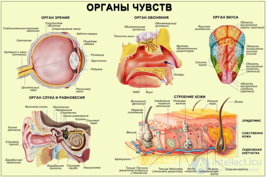
The sense organs are specialized organic structures located on the body and inside the organism, designed to perceive external information and to process and store it. They comprise:
1) receptors, which are located on its surface. They are designed to perceive stimuli of any nature and to convert them into nerve impulses;
2) neural pathways, specialized nerve fibers that conduct excitation acquired from various receptors to particular parts of the brain and back;
3) parts of the central nervous system (CNS) designed to process the incoming information (excitation) in order to produce a return response to the stimulus. The sense organs are otherwise called sensory organs, which are part of the general sensory system for perceiving incoming information.
According to I. P. Pavlov, a sensory system is a part of the nervous system consisting of a receptor apparatus that perceives internal or external stimuli, conducting neural pathways, and a part of the central nervous system that transforms the information arriving along them from the receptors.
The conducting neural pathways can be divided into:
1) afferent pathways, along which nervous excitation passes from the receptors to a specific part of the brain;
2) efferent pathways, along which a nerve impulse passes from the central nervous system to the periphery.
The totality of the afferent and efferent conducting pathways, including the receptors of a particular sense organ and the subcortical and cortical parts of the central nervous system that transform information, is called an analyzer.
A human being has five sense organs that establish his connection with the surrounding reality. They are divided into contact senses (through direct contact with the stimulus) and distance (distal) senses, which respond to remote stimuli:
1) contact senses: taste and touch;
2) distance senses: vision, hearing, and smell. The activity of each of the sense organs
constitutes an elementary mental process, sensation. Sensory information from external stimuli enters the central nervous system by two routes:
1) characteristic (specific) sensory pathways:
a) vision: through the retina, the lateral geniculate body, and the superior colliculi of the quadrigeminal plate to the primary and secondary visual cortex;
b) hearing: through the cochlear nuclei and the quadrigeminal nuclei, and the medial geniculate body to the primary auditory cortex;
c) taste: through the medulla oblongata and thalamus to the somatosensory cortex;
d) smell: through the olfactory bulb and the piriform cortex to the hypothalamus and the limbic system;
e) touch: passes through the spinal cord, brainstem, and thalamus to the somatosensory cortex;
2) nonspecific sensory pathways: pain and temperature sensations, located in the nuclei of the thalamus and brainstem.
Experimental confirmation of the proposition that sensation is a function of the cerebral cortex was given by I.P. Pavlov in his doctrine of analyzers.
An analyzer is a unified physiological system adapted to perceiving stimuli of the external or internal world, processing them into a nerve impulse, and forming a sensation and a general image of an object.
The following analyzers are distinguished: pain, vestibular, motor, visual, interoceptive, skin, olfactory, auditory, temperature, and others. Every analyzer has a fundamentally identical structure (Fig. 5.6). It consists of three parts.
1. The perceiving part of the analyzer is represented by receptors. They developed in the course of evolution as a result of the heightened sensitivity of certain cells to a particular kind of energy (thermal, chemical, mechanical, etc.). The stimulus to which a receptor is specifically adapted is called adequate, and all others are inadequate. Depending on their location
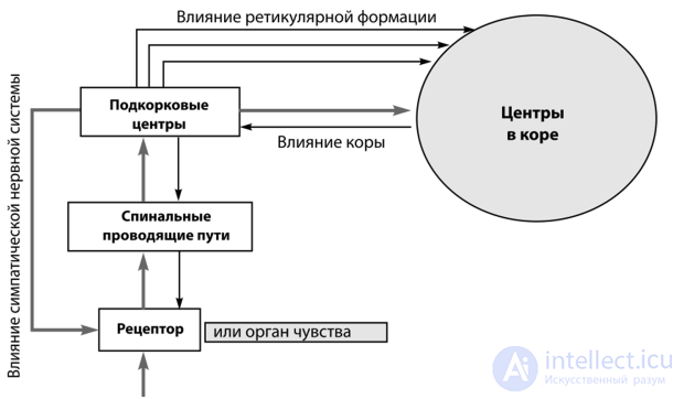
Fig. 5.6. The general principle of the structure of analyzers (R.I. Aizman et al., 2010), the following are distinguished. Exteroceptors (visual, auditory, olfactory, gustatory, tactile) lie on the surface of the body and respond to external influences, thereby ensuring an inflow of sensory information from the external environment. Interoceptors are located in the tissues of the internal organs (for example, chemoreceptors) and in the lumen of large vessels (baroreceptors), and are sensitive to various parameters of the internal environment (the concentration of chemically active substances, blood pressure, etc.); they are important for obtaining information about the functional state of the organism and its internal environment. Proprioceptors lie in the muscles, tendons, and joint capsules and perceive information about the degree of stretching and contraction of the muscles, thanks to which the "body sense" (the sensation of one's own body) and the perception of the relative position of its parts are formed.
The main part of the sense organs is the receptors (visual, auditory), which perceive the adequate stimulus. The function of receptors is to perceive energy of a certain kind and convert it into nerve impulses of a certain frequency (the sensory code). Therefore any receptor consists of two parts: the perceiving part proper and the transforming part. The first is represented by specific cells or other simple or complex structures adapted to interact with the stimulus. This part of the receptor is specific and interacts only with a certain kind of energy. The transforming part is the same in all receptors and is represented by the ending of a sensory nerve. In this part a stream of nerve impulses is formed, which is directed to the CNS.
2. The conduction section of each analyzer is represented by a sensory nerve along which excitation passes from the receptors to the subcortical and cortical centers of that analyzer. Two pathways, linked by collaterals, are distinguished here. The first, the so-called specific pathway of the analyzer, runs through the specific nuclei of the brainstem and plays the main role in transmitting sensory information and in the emergence of sensations of a particular modality. The second, the nonspecific pathway, is represented by neurons of the reticular formation; the stream of impulses passing along it changes the functional state of the structures of the spinal cord and brain, that is, it exerts a regulatory influence. It is well known that the reticular formation raises the activity of the cerebral cortex and creates a higher level of its excitation, which contributes to an increase in the intensity of the resulting sensation. The activity of the nonspecific system itself is maintained because nerve impulses constantly travel from various receptors along the specific pathway and reach the reticular formation through collaterals. The role of the conduction section of each analyzer is not limited to transmitting excitation from the receptors to the cortex: it also participates in the emergence of sensations. For example, the subcortical centers of the visual analyzer, located in the midbrain (in the superior colliculi), receive information from the visual receptors and change the curvature of the lens and the width of the pupil, thereby tuning the organ of vision to a more accurate perception of visual information. In addition, vague, crude sensations already arise at the level of the diencephalon (for example, sensations of light and shadow, of light and dark objects, are formed in the centers of the lateral geniculate bodies). Considering the conduction part of the analyzers as a whole, attention should be paid to the thalamus. In this part of the diencephalon the afferent pathways of all analyzers (with the exception of the olfactory one) converge. This means that the thalamus receives information from the extero-, proprio-, and interoceptors about the surrounding environment and the state of the organism.
About forty nuclei participate in processing impulses of all kinds of sensitivity. There are four types of thalamic nuclei. The first type is the specific relay nuclei of the various sensory systems. Information of a particular modality (visual, gustatory, tactile, pain, etc.) arrives here from the corresponding receptive fields. After being switched here, it is clearly projected onto the corresponding sensory zone of the cortex, or more precisely, onto the primary field of that zone. The specific nuclei do not merely perform switching, as was originally thought (the relay function), but also analyze the specialized information concentrated there, separating the essential from the inessential.
The second type of thalamic nuclei is the nonspecific nuclei. Sensory information arriving here is switched to the neurons of the reticular formation (that is, the nonspecific pathways of the analyzers pass through here). The nonspecific nuclei are connected with all areas of the cortex, as well as with the hypothalamus (the highest subcortical center regulating autonomic functions) and the limbic system. For this reason, various sensations evoke particular emotions and are also accompanied by changes in the functioning of the internal organs.
The third type is the association nuclei. Their very name (Latin association — connection, union, link) indicates that information of various modalities converges here, arriving via collaterals from the specific nuclei. The processes of integration and analysis taking place in the nuclei with association functions can apparently be regarded as the subcortical stage in the formation of holistic images. From here, information travels along ascending neurons to the secondary cortical fields (projection-association fields). And finally, the thalamus also contains motor nuclei, which have connections with the motor zones of the cortex.
Thus, all sensory information is gathered and analyzed in the thalamus. Here it is partly processed and, in this processed form, transmitted to various areas of the cortex. Most sensory information does not reach the highest division of the central nervous system (and therefore does not produce distinct, conscious sensations), but becomes a component of motor and emotional responses and, possibly, "material" for intuition.
3. The central division of each analyzer is represented by a particular area of the cerebral cortex (Fig. 5.7). Neurons go to the primary
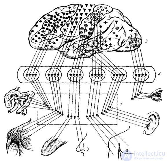
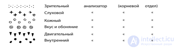
Fig. 5.7. Cortical localization of the central divisions of the analyzers (after K. Bykov, 1956; cited in A.V. Korobkov and S.A. Chesnokova, 1986):
7 — specific nuclei of the brainstem; 2 — specific nuclei of the thalamus; 3 — cortical centers of the analyzers fields directly from the specific nuclei of the thalamus; they are built on the topical principle, that is, they are a projection of each part of the receptive field ("point to point"), for which they are called projection fields. The primary fields provide for the emergence of specific sensations and the finest, most detailed analysis of individual parameters of the stimulus. The secondary fields of the analyzers are located around the primary ones and are connected with them anatomically and functionally. They have more complex connections with the subcortex. More processed information reaches them, since it comes not directly from the relay nuclei of the thalamus but through the association nuclei. The secondary fields have a well-developed third layer, in which neurons of one area of the cortex are switched to neurons of another, that is, the association function is provided. Owing to the synthesis of information, what arises here is not individual sensations but holistic sensory images. For example, in the secondary fields of the visual system there arise not merely sensations of light, of various colors and lines, but images of objects — faces, scenes of nature, and so on — and their recognition takes place. Since the secondary fields link the primary (projection) fields with the tertiary (association) fields, they are called projection-association fields.
The tertiary fields are the association fields. They are located between the nuclear zones of the various analyzers, provide complex interconnections among them, have no direct contacts with the subcortex, but are connected with it indirectly, through the secondary fields.
The development of higher mental functions is associated not only with the integration of sensory components with one another, but also of sensory components with motor ones. Each analyzer includes descending, efferent neurons that "switch on" motor reactions. For example, visual information arriving at the superior colliculi evokes "local" reflexes — involuntary eye movements following a moving object, one of the elements of the orienting reflex. In the cortex, the central ends of all analyzers are connected with the motor zone, which is located in the precentral gyrus and the premotor area and is the central division of the motor analyzer. Thus, the motor cortex receives information from all the afferent systems of the organism and serves as a connecting link in intersensory relations. In this way the connection between sensations and movements is ensured.
At present, in many foreign laboratories the doctrine of analyzers is called the physiology of general sensory systems. A sensory system consists of two parts: the objective sensory system, which includes the whole complex of physiological processes developing in all sensory systems, regardless of whether they occur in humans or in animals. The subjective sensory system is characteristic only of humans and is associated with the emergence of sensation or perception. In humans these systems are interconnected.
Thus, despite their specificity, all analyzers have a common structural scheme (receptor, conductive, and cortical divisions). This structure is characterized by:
Thus, the structural elements of the analyzers are not isolated in the nervous system but are anatomically and functionally connected with the speech centers, the limbic system, the ancient and old cortex, the autonomic centers of the brainstem, and so on, which ensures the interconnection of sensations with emotions, movements, behavior, and speech, and explains the influence of sensory information on the human organism.
Analyzers are figuratively called windows on the world, or channels of communication between a person and the external world and his or her own organism. Analysis of information already takes place "at the input," which is achieved through the selective responsiveness of the receptors. Not only the receptors but all the structural elements of each sensory system are adapted to the best possible detection and transmission of adequate signals, to which their sensitivity is highest. Thus, analyzers perform the function of signal detection.
Within a single modality there is an enormous variety of signals: sounds, for example, vary in pitch, timbre, and origin; visual information varies in color, brightness, shape, size, and so on. The ability to sense the difference between them at the physiological level is due to the fact that different sensory signals arise in the analyzers in response to different stimuli. This function is called signal discrimination. It is achieved by the formation, at the level of the receptors, of nerve impulses with different frequency characteristics (the sensory code) and by the involvement of different elements of the sensory system. The quantitative aspect of discriminating sensations is reflected by the difference threshold. The function of signal discrimination is carried out by all divisions of the analyzers, from the receptors to the cortex. In essence, it is an integral part of the process of analysis. As the child develops and his or her interaction with the external world becomes more complex, discriminations become increasingly fine. This is promoted by the development of each analyzer separately, as well as by the growing complexity of their interaction. Fine discrimination of complex signals at the cortical level is manifested in reduced generalization and strengthened differential inhibition. Movements play a major role in this process: motor discriminations help sensory ones. Thus, discriminating visual information requires eye movements, which inevitably accompany the process of looking at an object, as well as various positions of the hands arising when it is felt by touch. For the development of musical ear, that is, the ability to hear the slightest difference in pitch, it is necessary not only to perceive sounds by ear but also to reproduce them with one's own voice, in order to feel the difference in the position of the larynx and the degree of tension of the vocal cords. The same principle operates in the formation of phonemic hearing. To discriminate speech sounds — phonemes — well, it is not enough to hear another person's speech (even if the speaker's diction is excellent); one must also feel one's own articulatory apparatus well (lips, tongue, palate, larynx, cheeks) and sense the differences in its positions when producing sounds. Many methods of teaching preschool and primary school children, as well as remedial techniques, rely on this mechanism.
It should be noted that the differentiation of individual sensations and holistic images is not the result of an automatic engagement of the corresponding physiological mechanisms. Fine analysis of stimuli requires the activity of the knowing subject himself or herself. If a person wants to take part in a given activity and it evokes positive emotions (interest, joy), his or her sensory sensitivity to various signals increases considerably. Voluntary attention plays an active role in this process. The following example can serve as confirmation. It has been established experimentally that the activity of central and peripheral neurons of a sensory system depends not only on the presence of a specific stimulus acting on the receptors, but also on the behavior of the individual within whom the sensory process takes place. A change in behavior affects the degree to which various elements of the analyzer are involved in the work, even though the stimulus remains the same. This result is achieved through control, exerted by the cerebral cortex and the nearest subcortex over the lower divisions of the analyzers by means of efferent neurons.
Thus, sensory processes cannot be regarded entirely as the physiological correlate of the objective properties of objects, since they also reflect a subjective factor — the needs, emotions, and associated behavior of the subject, which influence the sensory images that arise.
The psychophysiological mechanism of discriminating even relatively simple signals cannot be considered solely within the confines of a single analyzer, since it is connected with the activation of the integrative processes of the brain, and consequently with consciousness, speech, and thinking.
One of the questions that arises in the study of sensory systems is how information is transmitted in the analyzers. In the receptors, under the influence of a stimulus, nerve impulses of a particular frequency are formed, which travel along the afferent pathways in groups — "volleys" or "bursts" (the sensory frequency code). It is believed that the number of impulses and their frequency constitute the language with which the brain records and transmits a message about the properties of the reflected object. In keeping with the spirit of the times, this process is called information coding.
Other methods of coding also exist. For example, depending on the strength of the stimulus, a different number of neural elements is engaged; when one or another quality changes, the localization of the excited elements may change. A particular feature of an object may excite a strictly defined neuron or group of neurons (positional coding). The movement of a stimulus across the receptor field is registered through the sequential engagement of different neural channels of the analyzer (different projection fibers), which leads to a shift in the areas of excitation and evokes the corresponding sensation of the movement of the object.
The nervous system registers not only the characteristics of the stimulus but also the temporal boundaries of its action, by means of special neurons that respond only to the onset of the stimulus (on-neurons) and its end (off-neurons), or to an abrupt change during the stimulus. For example, the visual system contains on-neurons, which respond to the switching on of light, off-neurons, which are excited at the moment of switching off, and also on-off neurons, which discharge in both cases.
At the present stage it is impossible to establish a clear correspondence between a particular property of a stimulus and the way it is registered in the nervous system. The existing scientific data describe only some principles of information transmission in the nervous system (Fig. 5.8).
In addition to such functions as signal detection, discrimination, transmission, and coding, sensory systems perform pattern recognition, which is conventionally considered the final operation of each analyzer. "To recognize" means to understand what it is or who it is, to realize the meaning of the sensory image that has arisen, to assign it to a particular class, to identify it with another image (and later, with a word).
The scheme of this process is as follows. Specific nerve impulses that have arrived from the receptors in the projection area of the cortex
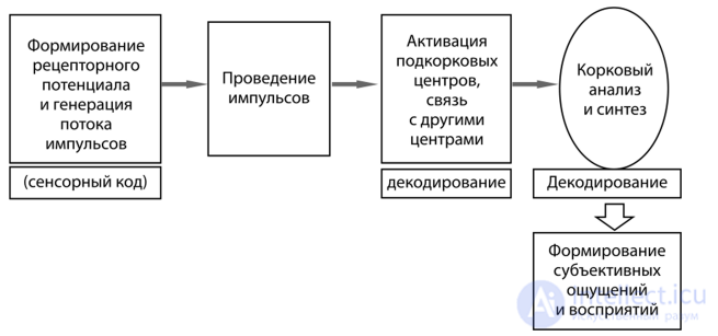
Fig. 5.8. General principle of the functioning of sensory systems (R.I. Aizman et al., 2010)
(in the primary field of the analyzer, where sensations are born) pass into the association fields (secondary and tertiary, where the synthesis of various sensations takes place), from there are directed to the hippocampus (memory) and structures of the limbic system (emotions), and then return to the cortex, reaching the projection field. Impulses from subcortical centers also arrive at the neurons of the projection cortex that retain traces of sensory excitation. The excitation is summed. The constructed sensory image is recognized and thereby becomes an act of consciousness through a physiological mechanism called information synthesis. At this moment the frontal cortex (movements) joins the process.
Thus, not only sensations but also movements, memory, and emotions participate in constructing a holistic image of an object and in recognizing it. Memory stores previously encountered impressions (sensory images), while emotions signal the significance of the information received.
If we consider the process of pattern recognition at the cellular level, we should turn our attention to the detector theory. Its name comes from the Latin words detectio — discovery, detector — discoverer. The point is that in all sensory systems, neurons have been found that respond only to some particular feature of their own. Detector neurons are arranged at all levels of the analyzer on a hierarchical principle. At the periphery of the analyzer lie neurons that recognize the simplest, individual features of the reflected object. From them, information converges on detector neurons for more complex properties of the reflected object, located at higher levels, and these in turn converge on those that reflect the holistic image.
The visual system has been studied best in this respect. Neurons have been found in it that are excited, for example, only by vertical or horizontal lines, by a particular angle of their inclination, by gaps in the contours of an object, by a particular color, or by the direction and speed of a moving object. More complex signals are also subject to detection. Thus, in the temporal lobe of primates (in its middle part) and of humans (in the temporo-occipital region), neurons have been found that "recognize" faces and individual features (eyes, nose, mouth). When this region is damaged, a person ceases to recognize familiar faces.
Perception does not arise mechanically or purely physiologically. The subject himself or herself, his or her consciousness and attention, takes an active part in its formation. In other words, the person must pay attention to the object, single it out, voluntarily shift attention from the whole to the parts, and have a desire and some goal for doing so. That is why teaching children can be successful only when it arouses in them a desire to learn what is offered to them, when it is of interest to them.

Comments