
Lecture 21 min.
DNA microarray, or DNA chip is a technology used in molecular biology and medicine. A DNA microarray consists of many small single-stranded molecules, DNA probes, which are covalently attached to a solid base . Each such probe has a strictly defined nucleotide sequence and a position on the microarray. Identical probes are located together, forming a microarray site. There is a one-to-one correspondence between a site and the DNA sequence of its probe. DNA microarrays are used to detect DNA or RNA (usually after reverse transcription), which may be either protein-coding or non-coding. Measuring gene expression by means of cDNA is called expression profiling, or expression analysis. On modern microarrays an entire genome can be placed, with every known gene serving as a probe .
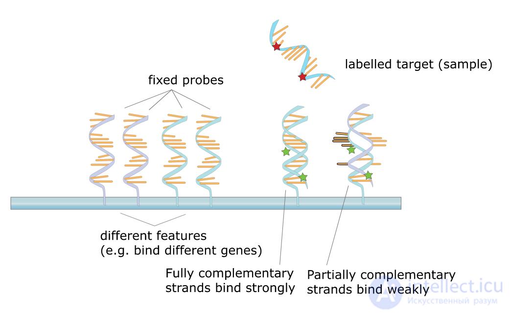
DNA microarrays are based on the phenomenon of hybridization. If only small amounts of DNA from the sample under study are available, amplification is carried out. For RNA, reverse transcription is performed first, although this is not required: there are chips that work with both DNA and RNA. Preparing DNA/RNA samples consists of labeling the samples with various fluorescent labels for later detection and applying the samples to the microarray. The DNA microarray with the sample applied is incubated for some time so that hybridization of complementary single-stranded molecules can occur, after which the chip is washed. All non-complementary DNA/RNA of the sample is washed off the chip. The microarray is then scanned with a laser, which causes the labeled sample molecules to fluoresce. A microscope connected to a computer measures the fluorescence of each site of the DNA microarray, and thereby determines the sequences of the hybridized DNA, which makes it possible to establish the sequence of the DNA or RNA in the sample .
DNA microarray technology originates from Southern blotting, a technique in which fragmented DNA is transferred to a suitable support and then, using a probe with a known nucleotide sequence, the content of the target sequence in the sample is determined. A set of different DNAs combined on a chip was first used in 1987 to determine features of the regulation of gene expression by interferons . Early DNA microarrays were made by "spotting" microquantities of cDNA onto filter paper. Miniature chips were used to determine features of gene expression in 1995 , and a complete eukaryotic genome (Saccharomyces cerevisiae) was placed on a microarray in 1997.
 Play media file
Amplified DNA fragments are applied with a micromanipulator to silicon or glass plates, with the probes fixed covalently. In 1991-1993 another approach was proposed, based on the photolithography technology used in the semiconductor industry. Later, in 1997, yet another method, "electrofocusing", was patented
Assembly begins with applying a special protective photosensitive layer, which makes the plate itself inert, to a glass plate measuring 12.8×12.8 mm. Only those places where the protective layer is illuminated are able to attach deoxyribonucleotides (A, T, G, C). After exposure to light, a solution of one of the bases is applied to the plate. All other places are shielded by a special photolithographic mask, so the deoxyribonucleotides are covalently attached to the plate in exactly the required places, after which everything that could not bind is washed away. The nucleotides themselves are chemically modified so that they can bind another nucleotide only when exposed to light . By repeating the cycles of mask, illumination, nucleotide application and washing as many times as necessary, and constantly changing the masks, unique sequences can be created. Different masks are designed to create a chip according to the requirements for the probes to be obtained .
Chains of identical single-stranded DNA, organized in a 90×90 μm square, grow on the surface of the plate nucleotide by nucleotide. Each square ultimately contains billions of identical probes. Photolithography is currently used to create relatively short probes, no longer than 100 nucleotides. The number of masks changed during the assembly of such a chip is comparable to the length of the probes: for example, assembling probes 18-25 nucleotides long on a chip requires about 40 masks. In practice, a large number of identical DNA microarrays are manufactured together on one large glass substrate .
Assembling this kind of microarray requires particularly complex substrates, which are essentially electronic chips with many outputs, each of which controls the voltage at a particular place on the plate. Unlike photolithography, where probes are assembled base by base, here ready-made single-stranded oligonucleotides are delivered to the required places on the plate under the action of an electric field. A positive voltage is created at the required place on the plate by means of the corresponding output of the microarray. Negatively charged single-stranded DNA moves toward the positive charge created and attaches at the required place. Then the next output of the microarray is activated, producing a positive charge at another place on the plate, to which the next probe is directed. The density of DNA on this kind of chip is much lower than on chips produced by photolithography .
There are three basic types of microarrays: for gene expression analysis (GEM microarray), for comparative genomic hybridization (MCGH), and for detection of single nucleotide polymorphisms (SNPM) . The basic protocol can be presented as follows :
1. Isolation of DNA/RNA
At this stage either mRNA (GEM) or fragments of genomic DNA (MCGH, SNPM) are isolated from the samples of interest. This is now easily done with special commercial kits.
2. Labeling of DNA/RNA
This process begins with reverse transcription (if necessary). The target fragment is then amplified by polymerase chain reaction. During PCR, nucleotides with fluorescent labels are incorporated into the DNA fragments
3. Hybridization
Here the fluorescently labeled amplified samples are used as targets to search the microarray for complementary strands, that is, strands able to form stable double strands, or duplexes, according to the complementarity rule. As an example, the complementary sequences 5'-GCATGCAT-3’ and 3’-CGTACGTA-5’. Since one of the strands of the resulting duplex is labeled, the signal from such a duplex can be detected.
4. Washing
As soon as hybridization is finished, the chip is removed from the solution containing the labeled DNA samples. The chip itself is then thoroughly washed by defined procedures, using various buffer mixtures, centrifugation and so on[10].
5. Scanning
This process involves the use of optical scanning instruments able to detect photons of a precisely defined wavelength. Each site of the chip is illuminated with a beam of light of a certain wavelength, which activates the fluorescent label. The microarray is "illuminated" with argon lasers. The activated fluorescent label emits a photon of a slightly longer wavelength, which is registered by the instrument. The more photons the instrument captures in one exposure, the higher the luminescence intensity of that spot, and therefore the greater the number of duplexes formed there.
6. Data analysis
The data are an array of luminescence intensity values for each individual site of the microarray. Using mathematical comparison methods, it is possible to reliably identify the sites of the chip where hybridization occurred, and therefore to determine the DNA/RNA sequence in the sample
This is the most common application of DNA microarrays. RNA isolated from a cell culture undergoes reverse transcription, yielding labeled cDNA. Sometimes one more step of transcription from the cDNA is required (for chips that work with RNA), producing labeled cRNA. There are several different ways to introduce labels into the target molecule: incorporation of fluorescently labeled nucleotides during cDNA or cRNA synthesis; use of biotin-modified nucleotides, which are then stained with fluorescently labeled streptavidin; and use during synthesis of modified nucleotides to which a fluorescent label can then be added[14].
DNA or RNA labeled in this way is hybridized on the microarray and then washed off. A fluorescent signal is detected at each spot of the chip. In the case of biotinylated samples, after hybridization the DNA microarray is stained with streptavidin containing fluorescent labels. Fluorescence is excited by laser light and is usually registered with a scanning confocal microscope[15].
Microarrays are also used together with chromatin immunoprecipitation to determine the binding sites of transcription factors (TFs) . Formaldehyde is added to an extract of cellular DNA, which leads to the formation of covalent cross-links between DNA and proteins. The DNA is then fragmented. The desired TF is isolated from the mixture by affinity chromatography using antibodies or tags that have been inserted into that TF beforehand by genetic engineering methods. After purification, the DNA is freed from the TF, amplified, fluorescently labeled and used for hybridization on a microarray. This technique is widely known as "ChIP-chip"[18], or chromatin immunoprecipitation on a microarray, but it has limitations because TFs can bind far from the gene they regulate.
DNA microarrays are widely used to detect single nucleotide polymorphisms (SNPs). There are several different approaches[:
Allele discrimination
The microarrays carry short probes (25 nucleotides for Affymetrix microarrays) containing all variants of the SNP in the center, since this position has the strongest effect on hybridization quality. The fragmented, amplified, fluorescently labeled sample DNA is applied to the microarray, where the sample and probe hybridize. A strong signal is registered in places of complete complementarity of the molecules[19].
"Golden Gate" assay
This method is based on the polymerase chain reaction. Molecules complementary to the genomic DNA, carrying various SNP modifications at the 3'-end and different primers for the subsequent PCR at the 5'-end, are placed into a solution of genomic DNA; in addition, a molecule complementary to the other strand of the genomic DNA, on the other side of the SNP and carrying one more PCR primer at the 5'-end, is added. The polymerase will carry out synthesis only from the primer whose 3'-end matches the SNP. As a result, depending on which primer with the altered 3'-end undergoes PCR, that SNP is the one observed in the sample[20].
Primer extension
Here the probes are chosen so that they cover the entire stretch of DNA right up to the SNP, not including the polymorphism. Fragmented genomic DNA hybridizes with such a chip, after which polymerase and fluorescently labeled nucleotides with 4 different labels at the 3'-end are added to the solution. Such nucleotides can be attached by the polymerase to an existing chain, but cannot then attach the next nucleotide. As a result, the probes are extended by one nucleotide, which corresponds to the SNP[21].
Evaluation of the terminal nucleotide
This method is similar to the primer extension method, with the difference that the probes are located not on a flat substrate but on many small beads. The SNP is likewise recognized by the color of the label of the single added nucleotide[22].
Despite the advantages of DNA microarrays, they also have limitations in application. It is assumed that the signal intensity (luminescence) registered at a particular site of the microarray depends linearly on the amount of DNA that has hybridized, which is not always the case: because of hybridization kinetics, the signal level obtained at a given spot is not a linear function of the concentration of that DNA in the sample. Thus, the amount of DNA in a sample can be estimated accurately only within a certain range of initial DNA concentrations that can still provide a linear relationship. Estimates for relatively very high or very low initial DNA concentrations in the sample will be inaccurate[15].
In the complex genomes of eukaryotes, especially mammals, there are many homologous genes whose sequences are very similar, which imposes additional requirements on the design of microarray probes[23][24]. A probe designed for one gene A may also "catch" genes B, C and D that happen to be homologous to gene A, which will distort the final picture.
Another limitation is related to the size of the database of known genes. A probe corresponding to an unknown gene cannot be synthesized, and no interactions of such genes can be detected. This problem is especially relevant for prokaryotes, since the genomes of even closely related organisms can differ significantly. For example, in the bacterial species Aggregatibacter actinomycetemcomitans, the genomes of different strains can differ by 20% of genes and, consequently, microarrays designed for one strain will not be able to detect another
A protein microarray is a technology used in molecular biology. A protein microarray is a solid support (base) to which thousands of different proteins (antigens, antibodies, enzymes, etc.) are covalently attached at different points of the microarray. Each individual protein forms a region of its own high concentration on the microarray. This technology is very similar to DNA microarray technology, except that the probes are proteins rather than single-stranded DNA molecules.
Three types of protein microarrays are currently in relatively wide use: analytical, functional and reverse-phase microarrays.
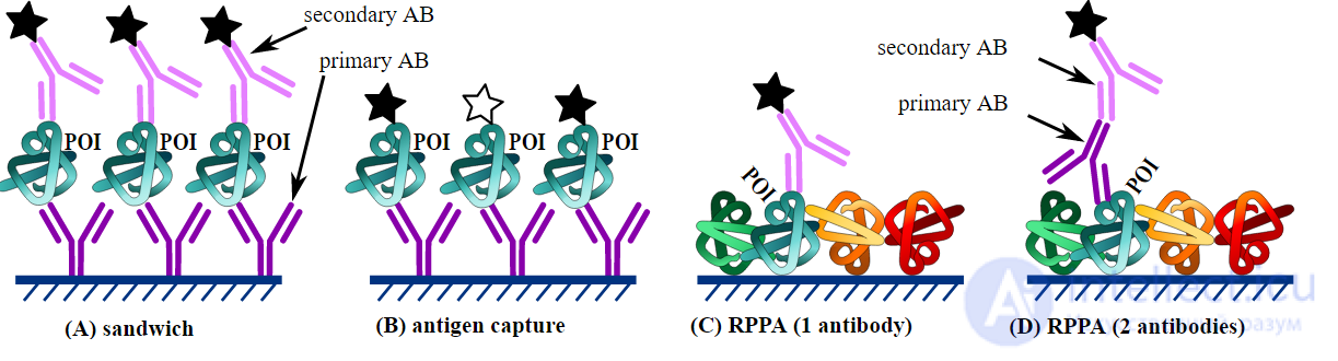
These microarrays are usually used to profile a complex mixture of proteins: measuring affinity, specificity and the expression level of a protein in the mixture. In this method, libraries of antibodies, aptamers and antigens are applied to solid supports that form the base of the microarray. The chip is then immersed in the protein mixture that is to be profiled. Antibody-based protein microarrays were the most widespread in the 2000s. Analytical microarrays can be used to search for differential expression and in clinical diagnostics. Examples include comparison of healthy and diseased tissues, as well as the response of a tissue to changes in environmental conditions
This type of microarray differs from analytical ones in that fully functional proteins or domains, rather than the analytical triad (antibodies, aptamers, antigens), are located on the solid base. Such microarrays make it possible to analyze the biochemical activity of an entire proteome in a single experiment. They are also used to study numerous interactions of proteins with other proteins, DNA, RNA, lipids and low-molecular-weight ligands
In making microarrays of this type, cells isolated from different tissues are lysed. The lysate is spotted onto a nitrocellulose membrane by a special machine, forming a microarray. Each spot of the resulting microarray is then analyzed with different antibodies against the protein of interest. This type of microarray makes it possible to detect proteins, or their conformational changes, that appear only during certain diseases
Analytical microarrays are also known as capture arrays. In this method, a library of antibodies, aptamers or affibodies is arrayed on the surface of a support. They are used as capture molecules, since each one binds specifically to a particular protein. The array is probed with a complex protein solution, such as a cell lysate.. Analysis of the resulting binding reactions using various detection systems can provide information on the expression levels of particular proteins in the sample, as well as measurements of binding affinity and specificity. This type of microarray is especially useful for comparing protein expression in different solutions. For example, the response of cells to a particular factor can be identified by comparing lysates of cells treated with specific substances or grown under specific conditions with lysates of control cells. Another application is the identification and profiling of diseased tissues. There are three types of protein microarrays that are currently used to study the biochemical activity of proteins.
Reverse-phase protein microarrays (RPPA) involve complex samples, such as tissue lysates. Cells are isolated from various tissues of interest and lysed. The lysate is spotted onto the microarray and probed with antibodies against the target protein of interest. These antibodies are usually detected by chemiluminescent, fluorescent or colorimetric assays. Reference peptides are printed on the slides so that the protein content of the lysate samples can be quantified. RPPAs make it possible to determine the presence of altered proteins or other agents that may result from disease. In particular, post-translational modifications, which are usually altered as a result of disease, can be detected using RPPAs
Functional protein microarrays (also known as target protein arrays) are constructed by immobilizing a large number of purified proteins and are used to identify protein-protein, protein-DNA, protein-RNA, protein-phospholipid and protein-small molecule interactions, to assess enzymatic activity, and to detect antibodies and demonstrate their specificity. They differ from analytical arrays in that functional protein arrays consist of arrays containing full-length functional proteins or protein domains. These protein chips are used to study the biochemical activity of an entire proteome in a single experiment.
A key element of any functional assay based on a protein microarray is that the proteins in the array must retain their native structure, so that meaningful functional interactions can take place on the surface of the array. The advantage of controlling the precise mode of attachment to the surface by means of a suitable affinity tag is that the immobilized proteins will have a homogeneous orientation, which leads to higher specific activity and a higher signal-to-noise ratio in the assays, with less interference from non-specific interactions. [
Detection methods for protein arrays must give a high signal and a low background. The most common and widely used detection method is fluorescent labeling, which is highly sensitive, safe, and compatible with readily available microarray laser scanners. Other labels can be used, such as affinity, photochemical or radioisotope labels. These labels are attached to the probe itself and can interfere with the reaction of the target protein. Therefore, a number of label-free detection methods are available, such as surface plasmon resonance (SPR), carbon nanotubes, carbon nanowire sensors (where detection occurs through a change in conductance), and microelectromechanical system (MEMS) cantilevers. [17]All of these label-free detection methods are relatively new and are not yet suitable for high-throughput detection of protein interactions; nevertheless, they hold a promising future. Immunoassays on micropillar scaffolds made of thiol-ene "synthetic paper" have been shown to generate an excellent fluorescence signal. [18]
Near-infrared fluorescence detection can be used for the quantification of protein on nitrocellulose-coated slides. This limits interference from nitrocellulose autofluorescence at the UV wavelengths used for standard fluorescence sensors. [19]
There are five main areas in which protein arrays are applied: diagnostics, proteomics, functional protein analysis, antibody characterization, and treatment development.
Diagnostics involves the detection of antigens and antibodies in blood samples; serum profiling to discover new disease biomarkers; monitoring of disease states and response to therapy in personalized medicine; and environmental and food monitoring. Digital bioassay is an example of the use of protein microarrays for diagnostic purposes. In this technology, an array of microwells on a glass/polymer chip is seeded with magnetic beads (coated with fluorescently labeled antibodies), exposed to target antigens, and then characterized under a microscope by counting the fluorescent wells. Recently, a cost-effective platform for fabricating such microwell arrays (using OSTE polymers) was demonstrated, and a model bioassay system was successfully characterized.[20]
Proteomics refers to the profiling of protein expression, that is, which proteins are expressed in the lysate of a particular cell.
Functional protein analysis is the identification of protein-protein interactions (for example, identifying members of a protein complex), protein-phospholipid interactions, small-molecule targets, enzymatic substrates (especially kinase substrates), and receptor ligands.
Antibody characterization characterizes cross-reactivity, specificity and epitope mapping.
Treatment development includes the development of antigen-specific therapies for autoimmune diseases, cancer and allergy; and the identification of small-molecule targets that could potentially be used as new drugs.
Despite significant investment by several companies, protein chips have not yet flooded the market. Manufacturers have found that proteins are actually quite difficult to work with. Producing reliable, consistent, high-throughput proteins that are correctly folded and functional is difficult, since it often results in low protein yields because of reduced solubility and the formation of inclusion bodies. [ citation needed ] Creating a protein chip requires many more steps than creating a DNA chip .
There are a number of approaches to this problem, which differ fundamentally depending on whether the proteins are immobilized through non-specific, poorly defined interactions or through a defined set of known interactions. The first approach is attractive for its simplicity and is compatible with purified proteins obtained from native or recombinant sources [21] [22], but suffers from a number of risks. The most notable of these relate to the uncontrolled nature of the interactions between each protein and the surface; at best this could give rise to a heterogeneous population of proteins in which the active sites are sometimes occluded by the surface; at worst it can destroy activity completely because of partial or complete surface-mediated unfolding of the immobilized protein.
The challenges include: 1) finding a surface and an attachment method that allows proteins to retain their secondary or tertiary structure, and hence their biological activity and their interactions with other molecules, 2) creating an array with a long shelf life, so that the proteins on the chip do not denature within a short time, 3) identifying and isolating antibodies or other capture molecules against every protein in the human genome, 4) quantifying the levels of bound protein while ensuring sensitivity and avoiding background noise, 5) extracting the detected protein from the chip for its further analysis, 6) reducing non-specific binding by the capture agents, 7) the capacity of the chip must be sufficient to image as complete a representation of the proteome as possible; abundant proteins overwhelm the detection of less abundant proteins, such as signaling molecules and receptors, which are usually of greater therapeutic interest.

Comments