
Lecture 34 min.
Optogenetics is a multifaceted research field in biology and neuroscience that combines genetic methods and optical technologies to control the activity of neurons and other cells using light. This field of research is of great importance for understanding the functions of the nervous system, as well as for developing treatments for various neurological and psychiatric disorders.
Optogenetics is a technique for studying the work of nerve cells, based on introducing into their membrane special channels — opsins — that respond to excitation by light. If the brain is exposed to light of a certain wavelength, the neurons that have such channels will be activated or, conversely, will be unable to generate action potentials.
Genetic engineering methods are used to express the channels, and lasers, optical fiber and other optical equipment are used for the subsequent activation or inhibition of neurons and neural networks.
Optogenetics emerged in 2005, and the first opsin used was channelrhodopsin-2 (ChR2) .
The advantage of optogenetic methods over traditional electrophysiological methods of studying neural networks and influencing them lies in the possibility of highly selective activation or suppression of specific neuronal connections . This selectivity opens up new possibilities in the therapy of Parkinson's disease, depression, anxiety and epilepsy .
The important components of optogenetics include:
Optogenetic tools: Optogenetics uses optogenetic tools such as rhodopsins and chloride channels, which can be introduced into neurons using genetic engineering. These tools respond to light of various wavelengths and can change the membrane potentials of neurons.
Genetic engineering: To introduce optogenetic tools into cells, genetic engineering methods are used, such as viral transduction or transgenic models, in order to create living organisms with integrated optogenetic proteins.
Lasers and light sources: Light is required to activate optogenetic tools. Lasers and optical fibers are used for the precise and controlled delivery of light to specific regions of the organism.
The applications of optogenetics are diverse. They include studies of brain activity, including the study of the mechanisms of memory and learning, as well as the search for approaches to treating diseases associated with neurological disorders, for example depression, schizophrenia and Parkinson's disease.
Optogenetics makes it possible to manipulate the activity of cells with high precision, which makes it a powerful tool in neuroscience research and in the development of new treatments for neurological disorders.
In 1979, Francis Crick suggested that controlling all cells of one type in the brain, while leaving the others more or less unchanged, was a real challenge for neuroscience. Francis Crick suggested that a technology using light might be useful for controlling neuronal activity with temporal and spatial precision, but at that time there was no technique that could make neurons respond to light.
By the early 1990s, L. C. Katz and E. Callaway had shown that light could release glutamate. In 1994, Häberle and Bühldt had already demonstrated functional heterologous expression of bacteriorhodopsin for light-activated ion flow in yeast.
In 1995, Georg Nagel et al. and Ernst Bamberg attempted heterologous expression of microbial rhodopsins (also bacteriorhodopsin, and also in a non-neuronal system, Xenopus oocytes) ( Georg Nagel et al., 1995, FEBS Lett.) and demonstrated a light-induced current.
The earliest genetically targeted method that used light to control rhodopsin-sensitized neurons was described in January 2002 by Boris Zemelman and Gero Miesenböck , who used cultured mammalian neurons expressing Drosophila rhodopsin. In 2003, Zemelman and Miesenböck developed a second method of light-dependent activation of neurons, in which individual ionotropic channels TRPV1, TRPM8 and P2X2 were gated by photocaged ligands in response to light. Beginning in 2004, the groups of Kramer and Isacoff, in collaboration with the group of Trauner, developed organic photoswitches, or "reversibly caged" compounds , that could interact with genetically introduced ion channels. The TRPV1 technique, albeit without a light trigger, was subsequently used by several laboratories to alter feeding, locomotion and behavioral resilience in laboratory animals. However, light-based approaches to altering neuronal activity were not applied outside the original laboratories, probably because a more easy-to-use channelrhodopsin was cloned soon afterwards.
Peter Hegemann , studying the light response of green algae at the University of Regensburg, discovered photocurrents that were too fast to be explained by classical G-protein-coupled animal rhodopsins . Teaming up with the electrophysiologist Georg Nagel of the Max Planck Institute in Frankfurt, they were able to demonstrate that a single gene from the alga Chlamydomonas produces large photocurrents when expressed in a frog oocyte. To identify the expressing cells, they replaced the cytoplasmic tail of the algal protein with the fluorescent protein YFP , creating the first widely applicable optogenetic tool. In a 2003 paper they stated that "expression of ChR2 in oocytes or mammalian cells can be used as a powerful tool to increase the cytoplasmic concentration of Ca 2+ or to depolarize the cell membrane, simply by illumination".
Karl Deisseroth of the Department of Bioengineering at Stanford published the pages of his first experiment in early July 2004, showing light activation of neurons expressing channelrhodopsin. [30] In August 2005, members of his laboratory, including graduate students Ed Boyden and Feng Zhang , in collaboration with Georg Nagel , published the first demonstration of a single-component optogenetic system in neurons, using the channelrhodopsin-2(H134R)-eYFP mutant from Georg Nagel , which is the first mutant of channelrhodopsin-2 since its functional characterization by Georg Nagel and Hegemann.
Zhuo-Hua Pan of Wayne State University , investigating the possibility of restoring vision in blindness, tried targeting rhodopsin to ganglion cells — the neurons in our eyes that connect directly to the brain. According to Pan, his first observation of optical activation of retinal neurons by channelrhodopsin was in February 2004, [32] five months before Deisseroth's initial observation in July 2004 . Indeed, the transfected neurons became electrically active in response to light, and in 2005 Zhuo-Hua Pan reported successful in vivo transfection of channelrhodopsin into mouse retinal ganglion cells and electrical responses to photostimulation in retinal slice culture. [34] This approach was eventually implemented in a human patient by Botond Roska and colleagues in 2021.
In April 2005, Susana Lima and Miesenböck reported the first use of genetically targeted photostimulation of P2X2 to control animal behavior. They showed that photostimulation of genetically restricted groups of neurons, for example neurons of the dopaminergic system , elicits characteristic behavioral changes in fruit flies.
In October 2005, Lynn Landmesser and Stefan Herlitze also published the use of channelrhodopsin-2 to control neuronal activity in cultured hippocampal neurons and in spinal cord circuits of chick embryos developing intact. In addition, they were the first to introduce vertebrate rhodopsin, a light-activated G-protein-coupled receptor, as a tool for inhibiting neuronal activity by recruiting intracellular signaling pathways, likewise in hippocampal neurons and in the intact developing chick embryo.
The groups of Alexander Gottschalk and Georg Nagel created the first ChR2 mutant (H134R) and were the first to use channelrhodopsin-2 to control neuronal activity in intact animals, showing that motor patterns in the roundworm C. elegans can be evoked by light stimulation of genetically selected neural circuits (published in December 2005). In mice, controlled expression of optogenetic tools is often achieved with cell-type-specific Cre/loxP methods, developed for neuroscience by Joe Z. Tsien back in the 1990s [38] to activate or inhibit specific brain regions and cell types in vivo .
In 2007, the laboratories of Boyden and Deisseroth (together with the groups of Gottschalk and Georg Nagel ) simultaneously reported successful optogenetic inhibition of neuronal activity.
In 2007, the groups of Georg Nagel and Hegemann began optogenetic manipulation of cAMP. In 2014, Avelar et al. reported the first fungal rhodopsin-guanylyl cyclase gene. In 2015, Scheib et al. and Gao et al. characterized the activity of the rhodopsin-guanylyl cyclase gene. Both Shiqiang Gao et al. and Georg Nagel , Alexander Gottschalk identified it as the first 8-TM rhodopsin.
The powerful impact of optogenetic technologies on brain research has been recognized by numerous awards given to key players in the field.
In 2010, Georg Nagel , Peter Hegemann and Ernst Bamberg were awarded the Wiley Prize in Biomedical Sciences , and were also among the recipients of the Karl Heinz Beckurts Prize in 2010 . [45] That same year, Karl Deisseroth was awarded the first HFSP Nakasone Award for "pioneering work on the development of optogenetic methods for studying the function of neural networks underlying behavior."
In 2012, Bamberg, Deisseroth, Hegemann and Georg Nagel were awarded the Zülch Prize by the Max Planck Society , and Miesenböck was awarded the Baillet Latour Health Prize for "pioneering optogenetic approaches to manipulating neuronal activity and controlling animal behavior." "
In 2013, Georg Nagel and Hegemann were among the recipients of the Louis-Jeantet Prize for Medicine . Also in the same year, Bamberg, Boyden, Deisseroth, Hegemann, Miesenböck and Georg Nagel were jointly awarded the Brain Prize for "the invention and refinement of optogenetics."
In 2017, Deisseroth was awarded the Else Kröner Fresenius Research Prize "for discoveries in optogenetics and hydrogel-tissue chemistry, and for research on the neural circuits underlying depression."
In 2018, the Inamori Foundation presented Deisseroth with the Kyoto Prize for "leading optogenetics" and "revolutionary research in systems neuroscience."
In 2019, Bamberg, Boyden, Deisseroth, Hegemann, Miesenböck and Georg Nagel were awarded the Rumford Prize of the American Academy of Arts and Sciences in recognition of "their outstanding contributions related to the invention and refinement of optogenetics."
In 2020, Deisseroth was awarded the Heineken Prize for Medicine by the Royal Netherlands Academy of Arts and Sciences for the development of optogenetics and hydrogel-tissue chemistry.
In 2020, Miesenböck, Hegemann and Georg Nagel jointly received the Shaw Prize in Life Science and Medicine.
In 2021, Hegemann, Deisseroth and Dieter Oesterhelt received the Albert Lasker Basic Medical Research Award .
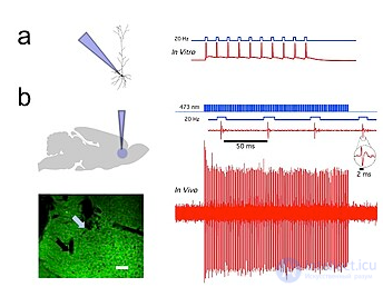
Fig. 1. Channelrhodopsin-2 (ChR2) induces temporally precise blue-light-evoked activity in rat prelimbic prefrontal cortical neurons. a) Schematic of the in vitro setup (left) showing blue light delivery and whole-cell patch-clamp recording of light-evoked activity from a fluorescent CaMKllα::ChR2-EYFP-expressing pyramidal neuron (right) in an acute brain slice. b) Schematic of the in vivo setup (left) showing blue light (473 nm) delivery and single-unit recording. (bottom left) Coronal brain slice showing CaMKllα::ChR2-EYFP expression in the prelimbic region. The blue arrow shows the tip of the optical fiber; the black arrow shows the tip of the recording electrode (left). White bar, 100 μm . (bottom right) Light recording of a prefrontal cortical neuron in vivo in a CaMKllα::ChR2-EYFP-transduced rat, showing light-evoked spiking in response to 20 Hz pulses of blue light (right). Inset: representative light-evoked single-unit response. [57]
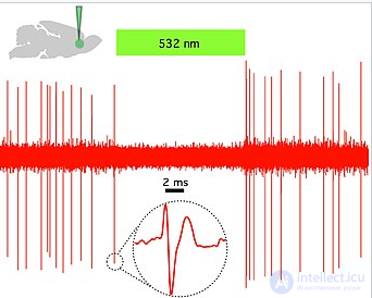
Fig. 2 . Halorhodopsin (NpHR) rapidly and reversibly silences spontaneous activity in vivo in the prelimbic prefrontal cortex of rats. (Top left) Schematic showing green light (532 nm) delivery in vivo and single-unit recording of a spontaneously active CaMKllα::eNpHR3.0-EYFP-expressing pyramidal neuron. (Right) Example trace showing that continuous 532 nm illumination inhibits single-unit activity in vivo . Inset: representative single-unit event; green bar, 10 seconds. [57]
Duration: 45 seconds.0:45
A nematode expressing the light-sensitive ion pump Mac. Mac is a proton pump originally isolated from the fungus Leptosphaeria maculans and now expressed in the muscle cells of C. elegans , which opens in response to green light and causes hyperpolarizing inhibition. Note the increase in the worm's body length each time it is exposed to green light, presumably caused by the muscle-relaxing effect of Mac. [58]
A nematode expressing ChR2 in a group of gubernacular oblique muscles responds to blue light stimulation. Blue light stimulation causes the gubernacular oblique muscles to contract repeatedly, producing repetitive thrusts of the spicule , as is naturally observed during copulation. [59]
Optogenetics provides temporal precision on the millisecond scale, which allows the experimenter to keep pace with the fast processing of biological information (for example, when investigating the causal role of specific action potential patterns in specific neurons). Indeed, to probe the neural code, optogenetics by definition must operate on the millisecond timescale in order to allow the addition or removal of precise patterns of activity within specific brain cells of intact animals, including mammals (see Figure 1 ) . By comparison, the temporal precision of traditional genetic manipulations (used to probe the causal role of specific genes within cells through "loss-of-function" or "gain-of-function" changes in those genes) is rather slow, from hours or days to months. It is also important to have fast readouts in optogenetics that can keep pace with the optical control. This can be done with electrical recordings ("optrodes") or with reporter proteins that are biosensors , where scientists couple fluorescent proteins to detector proteins. In addition, beyond its scientific impact, optogenetics is an important example of the value of both ecological conservation (since many of the key tools of optogenetics originate from microbial organisms occupying specialized ecological niches) and pure basic science, since these opsins were studied for decades by biophysicists and microbiologists for their own sake, without regard to their potential value for understanding neuroscience and neuropsychiatric diseases. [60]
Light-activated proteins: channels, pumps and enzymes
Thus, the hallmark of optogenetics is the introduction of fast, light-activated channels, pumps and enzymes that allow temporally precise manipulation of electrical and biochemical events while maintaining cell-type resolution through the use of specific targeting mechanisms. Among the microbial opsins that can be used to probe nervous system function are the channelrhodopsins (ChR2, ChR1, VChR1 and SFO) for neuronal excitation and anion-conducting channelrhodopsins for light-induced inhibition. Recently, potassium channels indirectly controlled by light have been engineered to prevent action potential generation in neurons during blue light illumination. [61] [62] Light-driven ion pumps are also used to inhibit neuronal activity, for example halorhodopsin (NpHR), [63] enhanced halorhodopsins (eNpHR2.0 and eNpHR3.0, see Figure 2), [64] archaerhodopsin (Arch ), fungal opsins (Mac) and enhanced bacteriorhodopsin (eBR).
Optogenetic control of well-defined biochemical events in behaving mammals is now also possible. Building on earlier work fusing vertebrate opsins to specific G-protein-coupled receptors [66], a family of chimeric single-component optogenetic tools was created that allowed researchers to manipulate, in behaving mammals, the concentration of specific intracellular messengers such as cAMP and IP3 in target cells. [67] Other biochemical approaches to optogenetics soon followed (especially with tools that showed low activity in the dark), when optical control of small GTPases and adenylyl cyclase was achieved in cultured cells using new strategies developed in several different laboratories. Photoactivated adenylyl cyclases were discovered in fungi and successfully used to control cAMP levels in mammalian neurons. This new repertoire of optogenetic actuators now allows cell-type-specific and temporally precise control of several axes of cellular function in intact animals.
Hardware for easy application
Another necessary factor is hardware (for example, integrated optical fiber and solid-state light sources) that allows control of specific cell types, even deep within the brain, in freely behaving animals. Most often the latter is now achieved using fiber-coupled diode technology, introduced in 2007, although, to avoid the use of implanted electrodes, researchers have developed ways to fit a zirconia "window" that was modified to become transparent and implanted into the skulls of mice, to allow optical waves to penetrate deeper and stimulate or suppress individual neurons. To stimulate superficial brain regions such as the cerebral cortex, optical fibers or LEDs can be attached directly to the animal's skull. To deliver light to deeper brain regions, more deeply implanted optical fibers are used. [78] In addition to fiber-optic approaches, fully wireless methods have been developed that use wireless power delivery to head-mounted LEDs for the unobstructed study of complex behavior in freely behaving organisms.
Expression of optogenetic actuators
Optogenetics also necessarily includes the development of genetic targeting strategies, such as cell-specific promoters or other tailored conditionally active viruses, to deliver light-sensitive probes to specific populations of neurons in the brains of living animals (for example, worms, fruit flies, mice, rats and monkeys). In invertebrates such as worms and fruit flies, some amount of all-trans retinal (ATR) is supplied with food. As noted above, a key advantage of microbial opsins is that they are fully functional in vertebrates without the addition of exogenous cofactors.
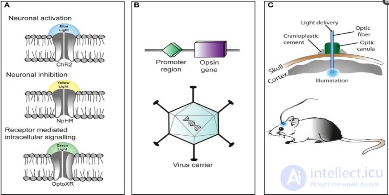
The three main components of applying optogenetics are the following: (A) Identification or synthesis of a light-sensitive protein (opsin), such as channelrhodopsin-2 (ChR2), halorhodopsin (NpHR), etc. (B) Design of a system for introducing genetic material containing the opsin into cells for protein expression, for example with Cre recombinase or the use of adeno-associated virus (C) light-emitting tools.
The optogenetic technique is flexible and adapts to the needs of the experimenter. Cation-selective channelrhodopsins (for example, ChR2) are used to excite neurons, while anion-conducting channelrhodopsins (for example, GtACR2) inhibit neuronal activity. Combining these tools in a single construct (for example, BiPOLES) allows both inhibition and excitation depending on the wavelength of illumination.
Introducing a microbial opsin into a specific subset of cells is challenging. A popular approach is to introduce an engineered viral vector containing the gene for an optogenetic actuator attached to a specific promoter , such as CAMKIIα , which is active in excitatory neurons. This provides some level of specificity, preventing, for example, expression in glial cells.
A more specific approach relies on transgenic "driver" mice that express Cre recombinase , an enzyme that catalyzes recombination between two lox-P sites, in a specific subset of cells, for example in interneurons expressing parvalbumin . When an engineered viral vector containing the gene for an optogenetic actuator between two lox-P sites is introduced, only cells producing Cre recombinase will express the microbial opsin. This method has made it possible to use several modified optogenetic actuators without having to create an entire transgenic animal line each time a new microbial opsin is needed.
After the microbial opsin has been introduced and expressed, a computer-controlled light source must be optically coupled to the brain region of interest. Light-emitting diodes (LEDs) or diode-pumped solid-state lasers (DPSS) delivered through optical fiber are often used. Recent advances include the emergence of wireless head-mounted devices that apply LEDs to target regions and, as a consequence, give animals more freedom of movement.
Fiber-based approaches can also be used to combine optical stimulation and calcium imaging . This allows researchers to image and manipulate the activity of individual neurons in awake animals. Simultaneous recording from several deep brain regions is also possible using GRIN lenses connected via an optical fiber to an external photodetector and photostimulator.
One of the main problems of optogenetics is that not all cells in question may express the microbial opsin gene at the same level. Thus, even illumination at a given light intensity will have different effects on individual cells. Optogenetic stimulation of brain neurons is even less controllable, because the intensity of light from the light source (for example, an implanted optical fiber) falls off exponentially.
It remains difficult to target opsins to specific subcellular compartments, such as the plasma membrane, synaptic vesicles or mitochondria. Restricting the opsin to specific regions of the plasma membrane, such as dendrites , somata or axon terminals , provides a clearer understanding of neural circuits.
Mathematical modeling shows that selective expression of opsin in specific cell types can radically alter the dynamic behavior of neural circuits. In particular, optogenetic stimulation that preferentially targets inhibitory cells can transform the excitability of neural tissue, also affecting non-transfected neurons.
The original channelrhodopsin-2 channel closed more slowly than typical cation channels of cortical neurons, which led to prolonged depolarization and calcium influx. Since then, many channelrhodopsin variants with more favorable kinetics have been created.
The difference between natural spiking patterns and optogenetic activation is that pulsed light stimulation causes synchronous activation of the expressing neurons, which rules out the possibility of sequential activity in the stimulated population. Consequently, it is difficult to understand how cells in the affected population communicate with each other or how their phase firing properties are related to circuit function.
Optogenetic activation has been combined with functional magnetic resonance imaging (fMRI) to elucidate the connectome, a detailed map of the brain's neural connections. [89] [92] Precisely timed optogenetic activation is used to calibrate the delayed hemodynamic signal ( BOLD ) on which fMRI is based.
The opsin proteins currently in use have absorption peaks across the visible spectrum, but remain highly sensitive to blue light. [89] This spectral overlap makes it very difficult to combine opsin activation with genetically encoded indicators ( GEVIs , GECIs , GluSnFR , synapto-pHluorin ), most of which require blue light excitation. Infrared-activated opsins would, at standard illumination levels, increase light penetration and improve resolution by reducing light scattering.
Because of scattering, a narrow beam of light stimulating neurons in a region of neural tissue can produce a response profile that is much wider than the stimulation beam. [93] In this case, neurons may be activated (or inhibited) unintentionally. Computer modeling tools [94] [95] are used to estimate the volume of stimulated tissue for different wavelengths of light.
The field of optogenetics has advanced the fundamental scientific understanding of how specific cell types contribute to the function of biological tissues such as neural circuits, in vivo . From a clinical perspective, optogenetics-based research has led to insights into Parkinson's disease and other neurological and psychiatric disorders such as autism , schizophrenia , drug abuse , anxiety and depression . Experimental treatment of blindness involves channelrhodopsin expressed in ganglion cells , which is stimulated by light patterns from specially designed goggles.
Optogenetic activation of olfactory sensory neurons was crucial for demonstrating the timing of odor processing [107] and the mechanism of neuromodulator-mediated olfactory behavior (for example , aggression , mating ) . In addition, optogenetics was used to replicate findings showing that the "afterimage" of odors is centered around the olfactory bulb, rather than at the periphery where the olfactory receptor neurons are located. Transgenic mice infected with Thy1-ChR2 channelrhodopsin were stimulated with a 473 nm laser positioned transcranially over the dorsal olfactory bulb. Longer photostimulation of mitral cells in the olfactory bulb led to observations of longer-lasting neuronal activity in this region after photostimulation ended, meaning that the olfactory sensory system is capable of undergoing long-term changes and recognizing differences between old and new odors.
Optogenetics, freely moving mammalian behavior, in vivo electrophysiology and slice physiology were integrated to investigate cholinergic interneurons of the nucleus accumbens by direct excitation or inhibition. Although these cholinergic cells make up less than 1% of the total accumbens neuron population, they are able to control the activity of dopaminergic terminals , which innervate medium spiny neurons (MSNs) in the nucleus accumbens. These accumbens MSNs are known to be involved in the neural pathways through which cocaine exerts its effects, since reducing cocaine-induced changes in the activity of these neurons has been shown to inhibit cocaine conditioning . The few cholinergic neurons present in the nucleus accumbens may prove to be viable targets for pharmacotherapy in the treatment of cocaine addiction.
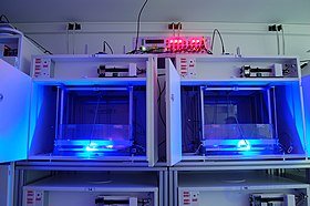
Rat cages equipped with optogenetic LED commutators, allowing in vivo study of animal behavior during optogenetic stimulation.
In vivo and in vitro recordings from the University of Colorado Boulder Optophysiology Laboratory of Donald C. Cooper, Ph.D. show individual pyramidal neurons expressing CAMKII AAV-ChR2 in the prefrontal cortex, which demonstrated high-fidelity action potential output with short 20 Hz pulses of blue light ( Fig. 1 ). [57]
Motor cortex
Repeated optogenetic stimulation in vivo in healthy animals could eventually cause seizures. [111] This model was named optokindling.
Piriform cortex
Repeated optogenetic stimulation of piriform cortex pyramidal cells in vivo in healthy animals could eventually cause seizures. [112] In vitro studies revealed a loss of feedback inhibition in the piriform circuit due to impaired GABA synthesis.
Optogenetics has been applied to atrial cardiomyocytes , to terminate spiral-wave arrhythmias that occur in atrial fibrillation , using light. [113] This method is still at the developmental stage. A recent study explored the potential of optogenetics as a method for correcting arrhythmias and resynchronizing cardiac pacing. In the study, channelrhodopsin-2 was introduced into cardiomyocytes of the ventricular regions of the hearts of transgenic mice, and in vitro photostimulation studies were performed on mice with both open and closed chests. Photostimulation led to increased cell activation and, thus, to stronger ventricular contractions, which led to an increased heart rate. In addition, this approach was applied in cardiac resynchronization therapy ( CRT ) as a new biological pacemaker, as a replacement for electrode-based CRT. [114] More recently, optogenetics has come to be used in the heart to defibrillate ventricular arrhythmias with local epicardial illumination, [115] generalized illumination of the whole heart [116] or customized stimulation patterns based on arrhythmogenic mechanisms, with the aim of reducing defibrillation energy. [117]
Optogenetic stimulation of the spiral ganglion in deaf mice restored auditory activity. [118] Optogenetic application to the cochlear region makes it possible to stimulate or inhibit spiral ganglion neurons (SGNs). In addition, because of the properties of SGN resting potentials, various variants of the channelrhodopsin-2 protein have been used, such as Chronos, [119] CatCh and f-Chrimson. [120] The Chronos and CatCh variants are particularly useful because they spend less time in the deactivated state, which allows greater activity with less blue light flash irradiation. In addition, the use of specially engineered red-shifted channels such as f-Chrimson allows stimulation with longer wavelengths, which reduces the potential long-term risk of phototoxicity without compromising gating speed. [121] As a result, the LED producing the light would require less energy, and the idea of a cochlear prosthesis combined with photostimulation becomes more feasible. [122]
Optogenetic stimulation of a modified red-light-excitable rhodopsin (ReaChR) expressed in the facial motor nucleus allowed minimally invasive activation of motor neurons that are effective in controlling whisker movements in mice. [123] In one new study, optogenetics was used in the dorsal raphe nucleus to activate and inhibit the release of dopaminergic substances in the ventral tegmental area. For activation, transgenic mice were infected with channelrhodopsin-2 with a TH-Cre promoter, and to achieve inhibition, the hyperpolarizing opsin NpHR was added to the TH-Cre promoter. The results showed that optical activation of dopaminergic neurons led to an increase in social interactions, while their inhibition reduced the need for social contact only after a period of isolation. [124]
Studying the visual system with optogenetics can be challenging. Indeed, the light used for optogenetic control can activate photoreceptors because of the proximity between the primary visual circuits and these photoreceptors. In this case, it is difficult to achieve spatial selectivity (especially in the case of the fly optic lobe). Thus, studying the visual system requires spectral separation using channels that are activated by wavelengths of light different from those of the rhodopsins inside photoreceptors (peak activation at 480 nm for rhodopsin 1 in Drosophila ). Red-shifted CsChrimson [125] or bistable Channelrhodopsin [126] are used for optogenetic activation of neurons (i.e. depolarization ), since both allow spectral separation. To achieve silencing of neurons (i.e. hyperpolarization ), an anion channelrhodopsin discovered in the cryptophyte alga species Guillardia theta (named GtACR1) can be used. [127] GtACR1 is more sensitive to light than other inhibitory tools such as halorhodopsin-class chloride pumps, and confers strong conductance. Because its activation peak (515 nm) is close to that of Rhodopsin 1, the optogenetic illumination as well as the visual stimulus must be carefully calibrated. Factors to consider are the wavelength of the optogenetic illumination (possibly higher than the GtACR1 activation peak), the size of the stimulus (to avoid activating the channels with the stimulus light) and the intensity of the optogenetic illumination. GtACR1 has been shown to be a useful inhibitory tool in optogenetic study of the Drosophila visual system by silencing the expression of T4/T5 neurons. [128] These studies can also be performed on intact, behaving animals, for example to investigate the optomotor response .
Optogenetic inhibition or activation of neurons tests their necessity and sufficiency, respectively, for the generation of behavior. [129] Using this approach, researchers can dissect the neural circuits controlling motor activity. By perturbing neurons at various locations in the sensorimotor system, researchers have learned about the role of descending neurons in the initiation of stereotyped behaviors, [130] how localized tactile sensory input [131] and the activity of interneurons [132] alter locomotion, and about the role of Purkinje cells in the generation and modulation of movement. [133] This is a powerful method for a broader understanding of the neural basis of animal locomotion and movement .
Currently available optogenetic actuators allow precise temporal control of the required intervention (i.e. inhibition or excitation of target neurons) with a precision that typically reaches the millisecond level. [134] However, temporal precision varies among optogenetic actuators [135] and depends on the frequency and intensity of stimulation. [93]
It is now possible to design experiments in which the light used for the intervention is triggered by a particular element of behavior (to suppress the behavior), a particular unconditioned stimulus (to associate something with that stimulus), or a particular oscillatory event in the brain (to suppress the event). A similar approach has already been used in several brain areas:
Sharp waves and ripple complexes (SWRs) are distinct high-frequency oscillatory events in the hippocampus , which are thought to play a role in memory formation and consolidation. These events can be easily detected by following the oscillatory cycles of the locally recorded online field potential . Thus, the onset of an event can be used as a trigger signal for a flash of light that is directed back into the hippocampus to inhibit neurons, especially during SWRs, as well as to optogenetically suppress the oscillations themselves. [138] Such "closed-loop" experiments are useful for studying SWR complexes and their role in memory.
Optogenetic control of cellular forces and induction of mechanotransduction. [139] The pictured cells are imaged over the course of an hour simultaneously with blue light that pulses every 60 seconds. This is also indicated by the flashing of a blue dot in the image. The cell relaxes for an hour without light activation, and then this cycle repeats again. The square inset magnifies the cell nucleus.
Similarly to how natural light-gated ion channels such as channelrhodopsin-2 enable optical control of ion flow, which is particularly useful in neuroscience, natural light-regulated signal transduction proteins also allow optical control of biochemical pathways, including both the generation of second messengers and protein-protein interactions, which is particularly useful in the study of cell biology and developmental biology. [140] In 2002, the first example was demonstrated of using photoproteins from another organism to control a biochemical pathway, using the light-induced interaction between plant phytochrome and phytochrome-interacting factor (PIF) to control gene transcription in yeast. By fusing phytochrome to a DNA-binding domain and PIF to a transcription activation domain, transcription of genes recognized by the DNA-binding domain could be induced by light. This study anticipated aspects of the later development of optogenetics in the brain, for example by suggesting that "directed delivery of light via fiber optics could potentially affect selected cells or tissues even within larger, opaque organisms". The literature is divided over whether the control of cell biochemistry with photoproteins should be included in the definition of optogenetics, since in common usage optogenetics refers specifically to the control of neuronal excitation with opsins, and since the control of neuronal excitation with opsins postdates and uses mechanisms different from the control of cell biochemistry with photoproteins. [140]
In addition to phytochromes, which are found in plants and cyanobacteria, LOV domains ( light-oxygen-voltage domain ) from plants and yeast and cryptochrome domains from plants are other natural photosensory domains that are used for optical control of biochemical pathways in cells. In addition, a synthetic photosensory domain based on the fluorescent protein Dronpa has been created for optical control of biochemical pathways. In photosensory domains, light absorption is coupled either to a change in protein-protein interactions (in the case of phytochromes, some LOV domains, cryptochromes and Dronpa mutants), or to a conformational change that exposes a linked protein segment or alters the activity of a linked protein domain (in the case of phytochromes and some LOV domains). [140] Light-regulated protein-protein interactions can then be used to recruit proteins to DNA, for example to induce gene transcription or DNA modifications, or to the plasma membrane, for example to activate resident signaling proteins. CRY2 also clusters when active, so it has been fused to signaling domains and subsequently photoactivated to enable clustering-based activation. [150] The LOV2 domain of Avena sativa (common oat) has been used to expose short peptides or an active protein domain in a light-dependent manner. Inserting this LOV domain into another protein can regulate function through light-induced peptide disruption. [154] The asLOV2 protein, which optogenetically exposes a peptide, has also been used as a scaffold for several synthetic systems of light-induced dimerization and light-induced dissociation (iLID and LOVTRAP, respectively). [155] [156] The systems can be used to control proteins through a protein-splitting strategy. [157] Photodissociable Dronpa domains have also been used to cage a protein's active site in the dark, release it after illumination with cyan light, and re-cage it after illumination with violet light. [158]
The ability to optically control signals for varying durations of time is being explored to elucidate how cell signaling pathways decode signal duration and respond with different outputs. [ Natural signaling cascades are able to respond with different outputs to differences in stimulus duration and dynamics. [160] For example, treating PC12 cells with epidermal growth factor (EGF, inducing a transient ERK activity profile) leads to cell proliferation, whereas the addition of nerve growth factor (NGF, inducing a sustained ERK activity profile) leads to differentiation into neuron-like cells. [161] This behavior was originally characterized using EGF and NGF applications, but the results were partially replicated using optical inputs. [162] In addition, using pulsed activation of a photoswitchable RAF engineered with photodissociable Dronpa domains, a rapid negative feedback loop was discovered in the RAF-MEK-ERK pathway. [158]
The research group of Professor Elias Manhares introduced optogenetic noise photostimulation. This is a method that uses random noisy light to activate neurons expressing ChR2. An optimal level of opto-noise photostimulation of the brain increases somatosensory evoked field potentials, the spiking frequency response of pyramidal neurons to somatosensory stimulation, and the amplitude of sodium current.

Comments