
Lecture
According to modern concepts, the nerve cell (neuron)
is the basic functional element
of the nervous system, which processes the information
arriving at it and passes the result of processing
on to other neurons [19].
1.1. Structure of the neuron
1.1.1. Soma, dendrites, axon
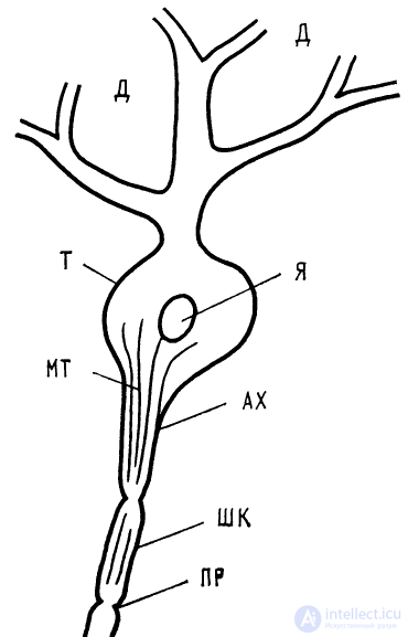
Fig. 1.1. Diagram of a neuron
D — dendrites, B — neuron body, N — nucleus, MT — microtubules, AH — axon hillock, SC — Schwann cells, NR — nodes of Ranvier
A neuron consists of a cell body — the soma, a set of
branching processes — the dendrites, through which
signals arrive, and usually a single
output fiber —
the axon. The intracellular
medium is separated from the
extracellular one by a thin
envelope — the membrane. On the
membrane of the soma and dendrites
there are synapses —
the endings of the axons
of other neurons (sometimes
of the neuron's own axon as well). The soma of the
neuron consists of the nucleus,
which contains the genetic
apparatus, and the cytoplasm,
in which protein
synthesis takes place. In the neuron body, in the dendrites and in the axons there are microtubules
about 200 Å in diameter, which presumably serve
to transport the proteins synthesized in the neuron body.
The transition of the neuron's soma into the axon is called the axon
hillock and is characterized by high excitability.
The axons of some neurons of the nervous system are covered
with sheaths consisting of Schwann cells or glial
cells. The gaps between them are the nodes of Ranvier
(Fig. 1.1).
Different types of nerve cells are characterized by different
geometric structures of the dendrites. There are
fairly long dendrites, extending from the soma
to a distance of 30–40 soma diameters, with little branching.
Other dendrites extend to a distance of about one diameter of the
soma and have a large number of branching nodes.
1.1.2. Structure of the neuronal membrane
The membrane, 70–80 Å thick, which separates the cytoplasm
of the neuron from the external environment, consists of a layer of phospholipid
molecules that has, on its outer and inner sides,
layers of protein molecules. The central phospholipid
layer has a symmetric structure in which the
fatty acids of the lipids face the inside of the membrane.
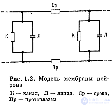
The membrane is pierced across by channels
through which certain ions can
move. Receptor proteins that control the state of the ion
channels are built into the membrane (Fig. 1.2). In the model of the membrane, the set of
channels is represented by a resistance, and the set of
dielectric regions by a capacitance connected in parallel.
The membrane surrounding the nerve cell (the soma,
dendrites and axon) is of two types: passive and active.
The passive membrane is able to conduct electrical
excitation only electrotonically (with attenuation), like a
cable. The active membrane is able to generate and
conduct an impulse that does not decay with distance. These two
types of membrane can alternate.
It has been established that the active membrane contains
special regions — channels — that can be in an open
or closed state. Channels are usually of two
types: those conducting sodium ions and those conducting potassium ions.
Channels of each of these types conduct only ions
of one kind (either only potassium or only sodium). The con-
trol (opening and
closing) of the channels
is carried out by polyvalent
cations, mainly
calcium. In an unexcited
membrane, calcium cations are in a bound
state with the molecules
that form the channels,
keeping the channels closed. A displacement
of the polar regions
of certain membrane molecules
opens the channels for
sodium and potassium ions, which
leads to excitation of the
membrane and generation
of a propagating
action potential.
1.1.3. Synapses, types of synapses
Synapses — regions of interaction of axon termi-
nals with the membrane of a neuron — are of two types:
excitatory and inhibitory. Excitatory synapses increase the
depolarization of the membrane, inhibitory ones — the hyperpolarization
of the membrane.
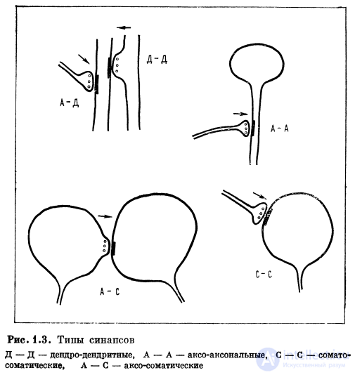
By the principle of their influence on the membrane, synapses
are divided into chemical and electrical. In
addition, axo-axonal, axo-somatic, axo-
dendritic, dendro-dendritic and somato-so-
matic synapses are distinguished (Fig. 1.3).
Although the density of synapses on the soma and
dendrites is on average the same, a higher
density of synapses occurs at the branching nodes
and in the regions of local dilations of the dendrites.
Therefore the branching nodes of the dendrites are of particular importance in the
functioning of the neuron.
On the soma of nerve cells the synapses are in most cases
small; their linear dimensions do not exceed 0.2–
0.4 µm (for the mammalian nervous system), whereas
on the dendrites a considerable part of the synapses has a
length of about 1 µm.
On the dendrites there are synaptic contacts of a
special kind, the so-called "spine" synapses,
located on mushroom-shaped processes. A spine consists of a thin stalk, whose end is thickened
3–5 times, forming the head of the spine. One or several ordinary synaptic
contacts are located on the head.
In some parts of the brain, the overwhelming majority of synapses
are located on spines.

Comments